In silico-designed lignin peroxidase fromPhanerochaete chrysosporiumshows enhanced acid stability for depolymerization of lignin.
Pham, L.T.M., Seo, H., Kim, K.J., Kim, Y.H.(2018) Biotechnol Biofuels 11: 325-325
- PubMed: 30555531 Search on PubMedSearch on PubMed Central
- DOI: https://doi.org/10.1186/s13068-018-1324-4
- Primary Citation Related Structures:
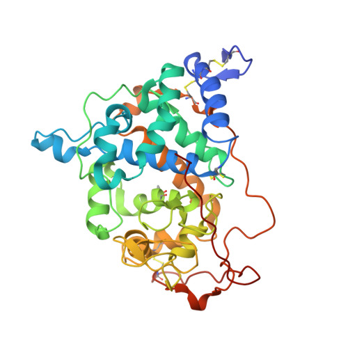
6A6Q - PubMed Abstract:
The lignin peroxidase isozyme H8 from the white-rot fungus Phanerochaete chrysosporium (LiPH8) demonstrates a high redox potential and can efficiently catalyze the oxidation of veratryl alcohol, as well as the degradation of recalcitrant lignin. However, native LiPH8 is unstable under acidic pH conditions. This characteristic is a barrier to lignin depolymerization, as repolymerization of phenolic products occurs simultaneously at neutral pH. Because repolymerization of phenolics is repressed at acidic pH, a highly acid-stable LiPH8 could accelerate the selective depolymerization of recalcitrant lignin. The engineered LiPH8 was in silico designed through the structural superimposition of surface-active site-harboring LiPH8 from Phanerochaete chrysosporium and acid-stable manganese peroxidase isozyme 6 (MnP6) from Ceriporiopsis subvermispora. Effective salt bridges were probed by molecular dynamics simulation and changes to Gibbs free energy following mutagenesis were predicted, suggesting promising variants with higher stability under extremely acidic conditions. The rationally designed variant, A55R/N156E-H239E, demonstrated a 12.5-fold increased half-life under extremely acidic conditions, 9.9-fold increased catalytic efficiency toward veratryl alcohol, and a 7.8-fold enhanced lignin model dimer conversion efficiency compared to those of native LiPH8. Furthermore, the two constructed salt bridges in the variant A55R/N156E-H239E were experimentally confirmed to be identical to the intentionally designed LiPH8 variant using X-ray crystallography (PDB ID: 6A6Q). Introduction of strong ionic salt bridges based on computational design resulted in a LiPH8 variant with markedly improved stability, as well as higher activity under acidic pH conditions. Thus, LiPH8, showing high acid stability, will be a crucial player in biomass valorization using selective depolymerization of lignin.
- 1School of Energy and Chemical Engineering, UNIST, 50 UNIST-gil, Ulju-gun, Ulsan, 44919 Republic of Korea.
Organizational Affiliation: