Amyloid fibril structure of alpha-synuclein determined by cryo-electron microscopy
Li, Y., Zhao, C., Luo, F., Liu, Z., Gui, X., Luo, Z., Zhang, X., Li, D., Liu, C., Li, X.(2018) Cell Res 28: 897-903
- PubMed: 30065316 Search on PubMedSearch on PubMed Central
- DOI: https://doi.org/10.1038/s41422-018-0075-x
- Primary Citation Related Structures:
6A6B - PubMed Abstract:
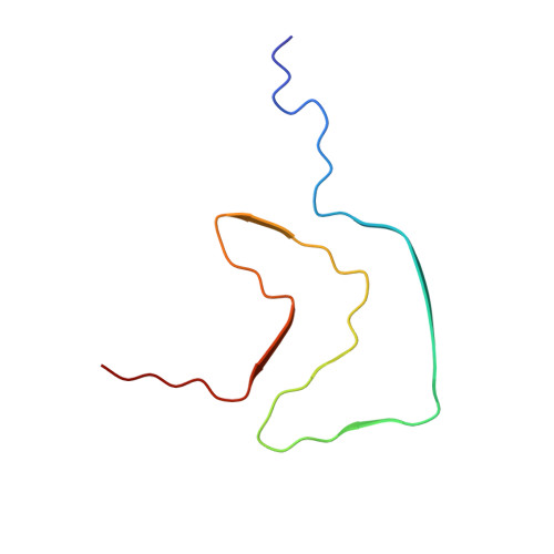
α-Synuclein (α-syn) amyloid fibrils are the major component of Lewy bodies, which are the pathological hallmark of Parkinson's disease (PD) and other synucleinopathies. High-resolution structure of α-syn fibril is important for understanding its assembly and pathological mechanism. Here, we determined a fibril structure of full-length α-syn (1-140) at the resolution of 3.07 Å by cryo-electron microscopy (cryo-EM). The fibrils are cytotoxic, and transmissible to induce endogenous α-syn aggregation in primary neurons. Based on the reconstructed cryo-EM density map, we were able to unambiguously build the fibril structure comprising residues 37-99. The α-syn amyloid fibril structure shows two protofilaments intertwining along an approximate 2 1 screw axis into a left-handed helix. Each protofilament features a Greek key-like topology. Remarkably, five out of the six early-onset PD familial mutations are located at the dimer interface of the fibril (H50Q, G51D, and A53T/E) or involved in the stabilization of the protofilament (E46K). Furthermore, these PD mutations lead to the formation of fibrils with polymorphic structures distinct from that of the wild-type. Our study provides molecular insight into the fibrillar assembly of α-syn at the atomic level and sheds light on the molecular pathogenesis caused by familial PD mutations of α-syn.
- Key Laboratory of Protein Sciences (Tsinghua University), Ministry of Education, School of Life Sciences, Tsinghua University, Beijing, 100084, China.
Organizational Affiliation: