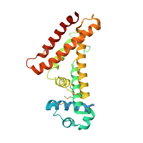
6A4L

AcrR from Mycobacterium tuberculosis


Experimental Data Snapshot

  • Method: X-RAY DIFFRACTION
  • Resolution: 2.80 Å
  • R-Value Free: 0.259 
  • R-Value Work: 0.213 
  • R-Value Observed: 0.215 

wwPDB Validation   3D Report Full Report


This is version 1.2 of the entry. See complete history


Literature

The crystal structure of AcrR from Mycobacterium tuberculosis reveals a one-component transcriptional regulation mechanism.

Kang, S.M.Kim, D.H.Jin, C.Ahn, H.C.Lee, B.J.

(2019) FEBS Open Bio 9: 1713-1725

  • DOI: https://doi.org/10.1002/2211-5463.12710
  • Primary Citation of Related Structures:  
    6A4L, 6A4W

  • PubMed Abstract: 

    Transcriptional regulator proteins are closely involved in essential survival strategies in bacteria. AcrR is a one-component allosteric repressor of the genes associated with lipid transport and antibiotic resistance. When fatty acid ligands bind to the C-terminal ligand-binding cavity of AcrR, a conformational change in the N-terminal operator-binding region of AcrR is triggered, which releases the repressed DNA and initiates transcription. This paper focuses on the structural transition mechanism of AcrR of Mycobacterium tuberculosis upon DNA and ligand binding. AcrR loses its structural integrity upon ligand-mediated structural alteration and bends toward the promoter DNA in a more compact form, initiating a rotational motion. Our functional characterization of AcrR and description of the ligand- and DNA-recognition mechanism may facilitate the discovery of new therapies for tuberculosis.


  • Organizational Affiliation

    The Research Institute of Pharmaceutical Sciences, College of Pharmacy, Seoul National University, Seoul, Korea.


Macromolecules
Find similar proteins by:  (by identity cutoff)  |  3D Structure
Entity ID: 1
MoleculeChains Sequence LengthOrganismDetailsImage
TetR family transcriptional regulator
A, B
230Mycobacterium tuberculosisMutation(s): 0 
Gene Names: 
UniProt
Find proteins for O07229 (Mycobacterium tuberculosis (strain ATCC 25618 / H37Rv))
Explore O07229 
Go to UniProtKB:  O07229
Entity Groups  
Sequence Clusters30% Identity50% Identity70% Identity90% Identity95% Identity100% Identity
UniProt GroupO07229
Sequence Annotations
Expand
  • Reference Sequence
Small Molecules
Ligands 2 Unique
IDChains Name / Formula / InChI Key2D Diagram3D Interactions
SO4
Query on SO4

Download Ideal Coordinates CCD File 
C [auth A],
D [auth A],
G [auth B],
H [auth B],
I [auth B]
SULFATE ION
O4 S
QAOWNCQODCNURD-UHFFFAOYSA-L
NA
Query on NA

Download Ideal Coordinates CCD File 
E [auth A],
F [auth A],
J [auth B]
SODIUM ION
Na
FKNQFGJONOIPTF-UHFFFAOYSA-N
Modified Residues  1 Unique
IDChains TypeFormula2D DiagramParent
MSE
Query on MSE
A, B
L-PEPTIDE LINKINGC5 H11 N O2 SeMET
Experimental Data & Validation

Experimental Data

  • Method: X-RAY DIFFRACTION
  • Resolution: 2.80 Å
  • R-Value Free: 0.259 
  • R-Value Work: 0.213 
  • R-Value Observed: 0.215 
  • Space Group: P 61
Unit Cell:
Length ( Å )Angle ( ˚ )
a = 118.752α = 90
b = 118.752β = 90
c = 93.456γ = 120
Software Package:
Software NamePurpose
PHENIXrefinement
HKL-2000data reduction
HKL-2000data scaling
PHENIXphasing

Structure Validation

View Full Validation Report


Entry History & Funding Information

Deposition Data

  • Released Date: 2019-06-26 
  • Deposition Author(s): Kang, S.M.

Funding OrganizationLocationGrant Number
National Research Foundation (Korea)Korea, Republic Of2018R1A2A1A19018526

Revision History  (Full details and data files)

  • Version 1.0: 2019-06-26
    Type: Initial release
  • Version 1.1: 2020-04-29
    Changes: Database references, Structure summary
  • Version 1.2: 2024-10-16
    Changes: Data collection, Database references, Derived calculations, Structure summary