6A11 | pdb_00006a11

Mandelate oxidase mutant-Y128F with phenylpyruvic acid


Experimental Data Snapshot

  • Method: X-RAY DIFFRACTION
  • Resolution: 1.45 Å
  • R-Value Free: 
    0.191 (Depositor), 0.191 (DCC) 
  • R-Value Work: 
    0.168 (Depositor), 0.168 (DCC) 
  • R-Value Observed: 
    0.169 (Depositor) 

Starting Model: experimental
View more details

wwPDB Validation 3D Report Full Report

Validation slider image for 6A11

Ligand Structure Quality Assessment 


This is version 1.2 of the entry. See complete history

Literature

Biochemical and structural explorations of alpha-hydroxyacid oxidases reveal a four-electron oxidative decarboxylation reaction.

Yeh, H.W.Lin, K.H.Lyu, S.Y.Li, Y.S.Huang, C.M.Wang, Y.L.Shih, H.W.Hsu, N.S.Wu, C.J.Li, T.L.

(2019) Acta Crystallogr D Struct Biol 75: 733-742

  • DOI: https://doi.org/10.1107/S2059798319009574
  • Primary Citation Related Structures: 
    5ZZQ, 5ZZS, 5ZZX, 5ZZY, 6A00, 6A0D, 6A0G, 6A0M, 6A0O, 6A0Y, 6A11, 6A1A

  • PubMed Abstract: 

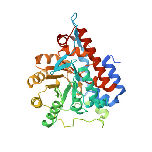
    p-Hydroxymandelate oxidase (Hmo) is a flavin mononucleotide (FMN)-dependent enzyme that oxidizes mandelate to benzoylformate. How the FMN-dependent oxidation is executed by Hmo remains unclear at the molecular level. A continuum of snapshots from crystal structures of Hmo and its mutants in complex with physiological/nonphysiological substrates, products and inhibitors provides a rationale for its substrate enantioselectivity/promiscuity, its active-site geometry/reactivity and its direct hydride-transfer mechanism. A single mutant, Y128F, that extends the two-electron oxidation reaction to a four-electron oxidative decarboxylation reaction was unexpectedly observed. Biochemical and structural approaches, including biochemistry, kinetics, stable isotope labeling and X-ray crystallography, were exploited to reach these conclusions and provide additional insights.


  • Organizational Affiliation
    • Genomics Research Center, Academia Sinica, Taipei 115, Taiwan.

Macromolecule Content 

  • Total Structure Weight: 40.65 kDa 
  • Atom Count: 3,077 
  • Modeled Residue Count: 334 
  • Deposited Residue Count: 377 
  • Unique protein chains: 1

Macromolecules

Find similar proteins by:|  3D Structure
Entity ID: 1
MoleculeChains  Sequence LengthOrganismDetailsImage
4-hydroxymandelate oxidase377Amycolatopsis orientalisMutation(s): 1 
Gene Names: hmo
EC: 1.1.3.46
UniProt
Find proteins for O52792 (Amycolatopsis orientalis)
Explore O52792 
Go to UniProtKB:  O52792
Entity Groups
Sequence Clusters30% Identity50% Identity70% Identity90% Identity95% Identity100% Identity
UniProt GroupO52792
Sequence Annotations
Expand
Reference Sequence

Small Molecules

Experimental Data & Validation

Experimental Data

  • Method: X-RAY DIFFRACTION
  • Resolution: 1.45 Å
  • R-Value Free:  0.191 (Depositor), 0.191 (DCC) 
  • R-Value Work:  0.168 (Depositor), 0.168 (DCC) 
  • R-Value Observed: 0.169 (Depositor) 
Space Group: I 4 2 2
Unit Cell:
Length ( Å )Angle ( ˚ )
a = 138.485α = 90
b = 138.485β = 90
c = 111.539γ = 90
Software Package:
Software NamePurpose
PHENIXrefinement
HKL-2000data reduction
HKL-2000data scaling
PHENIXphasing

Structure Validation

View Full Validation Report


Ligand Structure Quality Assessment 


Entry History 

Deposition Data

Revision History  (Full details and data files)

  • Version 1.0: 2019-06-19
    Type: Initial release
  • Version 1.1: 2019-08-14
    Changes: Data collection, Database references
  • Version 1.2: 2023-11-22
    Changes: Data collection, Database references, Refinement description