6A0I

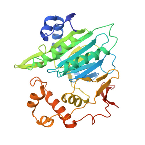
Crystal structure of human protein N-terminal asparagine amidohydrolase (NTAN1) C75S mutant


Experimental Data Snapshot

  • Method: X-RAY DIFFRACTION
  • Resolution: 2.00 Å
  • R-Value Free: 0.211 
  • R-Value Work: 0.164 
  • R-Value Observed: 0.166 

Starting Model: experimental
View more details

wwPDB Validation   3D Report Full Report


This is version 1.3 of the entry. See complete history


Literature

Structural Analyses on the Deamidation of N-Terminal Asn in the Human N-Degron Pathway.

Park, J.S.Lee, J.Y.Nguyen, Y.T.K.Kang, N.W.Oh, E.K.Jang, D.M.Kim, H.J.Kim, D.D.Han, B.W.

(2020) Biomolecules 10

  • DOI: https://doi.org/10.3390/biom10010163
  • Primary Citation of Related Structures:  
    6A0E, 6A0F, 6A0H, 6A0I

  • PubMed Abstract: 

    The N-degron pathway is a proteolytic system in which a single N-terminal amino acid acts as a determinant of protein degradation. Especially, degradation signaling of N-terminal asparagine (Nt-Asn) in eukaryotes is initiated from its deamidation by N-terminal asparagine amidohydrolase 1 (NTAN1) into aspartate. Here, we have elucidated structural principles of deamidation by human NTAN1. NTAN1 adopts the characteristic scaffold of CNF1/YfiH-like cysteine hydrolases that features an α-β-β sandwich structure and a catalytic triad comprising Cys, His, and Ser. In vitro deamidation assays using model peptide substrates with varying lengths and sequences showed that NTAN1 prefers hydrophobic residues at the second-position. The structures of NTAN1-peptide complexes further revealed that the recognition of Nt-Asn is sufficiently organized to produce high specificity, and the side chain of the second-position residue is accommodated in a hydrophobic pocket adjacent to the active site of NTAN1. Collectively, our structural and biochemical analyses of the substrate specificity of NTAN1 contribute to understanding the structural basis of all three amidases in the eukaryotic N-degron pathway.


  • Organizational Affiliation

    Research Institute of Pharmaceutical Sciences, College of Pharmacy, Seoul National University, Seoul 08826, Korea.


Macromolecules
Find similar proteins by:  (by identity cutoff)  |  3D Structure
Entity ID: 1
MoleculeChains Sequence LengthOrganismDetailsImage
Protein N-terminal asparagine amidohydrolase
A, B
318Homo sapiensMutation(s): 1 
Gene Names: NTAN1
EC: 3.5.1 (PDB Primary Data), 3.5.1.121 (UniProt)
UniProt & NIH Common Fund Data Resources
Find proteins for Q96AB6 (Homo sapiens)
Explore Q96AB6 
Go to UniProtKB:  Q96AB6
PHAROS:  Q96AB6
GTEx:  ENSG00000157045 
Entity Groups  
Sequence Clusters30% Identity50% Identity70% Identity90% Identity95% Identity100% Identity
UniProt GroupQ96AB6
Sequence Annotations
Expand
  • Reference Sequence
Small Molecules
Ligands 2 Unique
IDChains Name / Formula / InChI Key2D Diagram3D Interactions
PO4
Query on PO4

Download Ideal Coordinates CCD File 
AA [auth B],
O [auth A],
P [auth A]
PHOSPHATE ION
O4 P
NBIIXXVUZAFLBC-UHFFFAOYSA-K
GOL
Query on GOL

Download Ideal Coordinates CCD File 
C [auth A]
D [auth A]
E [auth A]
F [auth A]
G [auth A]
C [auth A],
D [auth A],
E [auth A],
F [auth A],
G [auth A],
H [auth A],
I [auth A],
J [auth A],
K [auth A],
L [auth A],
M [auth A],
N [auth A],
Q [auth B],
R [auth B],
S [auth B],
T [auth B],
U [auth B],
V [auth B],
W [auth B],
X [auth B],
Y [auth B],
Z [auth B]
GLYCEROL
C3 H8 O3
PEDCQBHIVMGVHV-UHFFFAOYSA-N
Experimental Data & Validation

Experimental Data

  • Method: X-RAY DIFFRACTION
  • Resolution: 2.00 Å
  • R-Value Free: 0.211 
  • R-Value Work: 0.164 
  • R-Value Observed: 0.166 
  • Space Group: P 21 21 21
Unit Cell:
Length ( Å )Angle ( ˚ )
a = 82.982α = 90
b = 84.34β = 90
c = 86.998γ = 90
Software Package:
Software NamePurpose
PHENIXrefinement
HKL-2000data reduction
HKL-2000data scaling
PHENIXphasing

Structure Validation

View Full Validation Report


Entry History & Funding Information

Deposition Data


Funding OrganizationLocationGrant Number
National Research Foundation (Korea)Korea, Republic Of2011-0030001
National Research Foundation (Korea)Korea, Republic Of2013M-3A6A-4043695

Revision History  (Full details and data files)

  • Version 1.0: 2019-12-11
    Type: Initial release
  • Version 1.1: 2020-06-24
    Changes: Database references
  • Version 1.2: 2023-11-22
    Changes: Data collection, Database references, Refinement description
  • Version 1.3: 2024-10-16
    Changes: Structure summary