5ZYX | pdb_00005zyx

Solution NMR structure of K30 peptide in 10 mM dioctanoyl phosphatidylglycerol (D8PG)


Experimental Data Snapshot

  • Method: SOLUTION NMR
  • Conformers Calculated: 100 
  • Conformers Submitted: 20 
  • Selection Criteria: structures with the lowest energy 

wwPDB Validation 3D Report Full Report

Validation slider image for 5ZYX

This is version 1.5 of the entry. See complete history

Literature

Design, Synthesis, Antibacterial Potential, and Structural Characterization of N-Acylated Derivatives of the Human Autophagy 16 Polypeptide.

Varnava, K.G.Mohid, S.A.Calligari, P.Stella, L.Reynison, J.Bhunia, A.Sarojini, V.

(2019) Bioconjug Chem 30: 1998-2010

  • DOI: https://doi.org/10.1021/acs.bioconjchem.9b00290
  • Primary Citation Related Structures: 
    5ZYX

  • PubMed Abstract: 

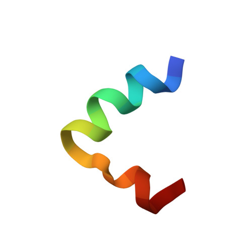
    A synthetic antimicrobial peptide library based on the human autophagy 16 polypeptide has been developed. Designed acetylated peptides bearing lipids of different chain lengths resulted in peptides with enhanced potency compared to the parent Atg16. A 21-residue fragment of Atg16 conjugated to 4-methylhexanoic acid ( K30 ) emerged as the most potent antibacterial, with negligible hemolysis. Several studies, including microscopy, dye leakage, and ITC, were conducted to gain insight into the antibacterial mechanism of action of the peptide. Visual inspection using both SEM and TEM revealed the membranolytic effect of the peptide on bacterial cells. The selectivity of the peptide against bacterial cell membranes was also proven using dye leakage assays. ITC analysis revealed the exothermic nature of the binding interaction of the peptide to D8PG micelles. The three-dimensional solution NMR structure of K30 in complex with dioctanoylphosphatidylglycerol (D8PG) micelles revealed that the peptide adopts a helix-loop-helix structure in the presence of anionic membrane lipids mimicking bacterial membranes. Intermolecular NOEs between the peptide and lipid deciphered the location of the peptide in the bound state, which was subsequently supported by the paramagnetic relaxation enhancement (PRE) NMR experiment. Collectively, these results describe the structure-function relationship of the peptide in the bacterial membrane.


  • Organizational Affiliation
    • School of Chemical Sciences , The University of Auckland , Private Bag 92019 , Auckland , New Zealand.

Macromolecule Content 

  • Total Structure Weight: 2.7 kDa 
  • Atom Count: 189 
  • Modeled Residue Count: 20 
  • Deposited Residue Count: 20 
  • Unique protein chains: 1

Macromolecules

Find similar proteins by:  Sequence   |   3D Structure  
Entity ID: 1
MoleculeChains  Sequence LengthOrganismDetailsImage
ARG-TRP-LYS-ARG-HIS-ILE-SER-GLU-GLN-LEU-ARG-ARG-ARG-ASP-ARG-LEU-GLN-ARG-GLN-ALA20Homo sapiensMutation(s): 0 
Membrane Entity: Yes 
UniProt & NIH Common Fund Data Resources
Find proteins for Q676U5 (Homo sapiens)
Explore Q676U5 
Go to UniProtKB:  Q676U5
PHAROS:  Q676U5
GTEx:  ENSG00000085978 
Entity Groups
Sequence Clusters30% Identity50% Identity70% Identity90% Identity95% Identity100% Identity
UniProt GroupQ676U5
Sequence Annotations
Expand
Reference Sequence

Experimental Data & Validation

Experimental Data

  • Method: SOLUTION NMR
  • Conformers Calculated: 100 
  • Conformers Submitted: 20 
  • Selection Criteria: structures with the lowest energy 

Structure Validation

View Full Validation Report


Entry History 

& Funding Information

Deposition Data


Funding OrganizationLocationGrant Number
India--

Revision History  (Full details and data files)

  • Version 1.0: 2019-06-12
    Type: Initial release
  • Version 1.1: 2019-06-19
    Changes: Data collection, Structure summary
  • Version 1.2: 2019-07-10
    Changes: Data collection, Database references
  • Version 1.3: 2019-07-31
    Changes: Data collection, Database references
  • Version 1.4: 2023-06-14
    Changes: Database references, Other
  • Version 1.5: 2024-05-15
    Changes: Data collection, Database references