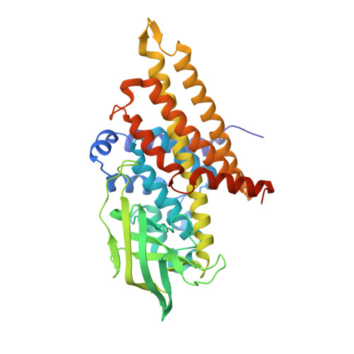
Crystal Structure of PigA: A Prolyl Thioester-Oxidizing Enzyme in Prodigiosin Biosynthesis.
Lee, C.-C., Ko, T.-P., Chen, C.T., Chan, Y.T., Lo, S.Y., Chang, J.Y., Chen, Y.W., Chung, T.F., Hsieh, H.J., Hsiao, C.D., Wang, A.H.J.(2019) Chembiochem 20: 193-202
- PubMed: 30095206
- DOI: https://doi.org/10.1002/cbic.201800409
- Primary Citation of Related Structures:
5ZW0, 5ZW2, 5ZW7, 5ZW8, 6AF6 - PubMed Abstract:
Prodigiosin is an intensely red pigment comprising three pyrroles. The biosynthetic pathway includes a two-step proline oxidation catalyzed by phosphatidylinositol N-acetylglucosaminyltransferase subunit A (PigA), with flavin adenine dinucleotide (FAD) as its cofactor. The enzyme is crystallized in the apo form and in complex with FAD and proline. As an acyl coenzyme A dehydrogenase (ACAD) family member, the protein folds into a β-sheet flanked by two α-helical domains. PigA forms a tetramer, which is consistent with analytical ultracentrifugation results. FAD binds to PigA in a similar way to that in the other enzymes of the ACAD family. The variable conformations of loop β4-β5 and helix αG correlate well with the structural flexibility required for substrate entrance to the Re side of FAD. Modeling with PigG, the acyl carrier protein, suggests a reasonable mode of interaction with PigA. The structure helps to explain the proline oxidation mechanism, in which Glu244 plays a central role by abstracting the substrate protons. It also reveals a plausible pocket for oxygen binding to the Si side of FAD.
- Institute of Biological Chemistry, Academia Sinica, 128 Academia Road Section 2, Taipei, 11529, Taiwan.
Organizational Affiliation:


