5ZU1 | pdb_00005zu1

Crystal Structure of BZ junction in diverse sequence


Experimental Data Snapshot

  • Method: X-RAY DIFFRACTION
  • Resolution: 3.01 Å
  • R-Value Free: 
    0.280 (Depositor), 0.279 (DCC) 
  • R-Value Work: 
    0.233 (Depositor), 0.232 (DCC) 
  • R-Value Observed: 
    0.238 (Depositor) 

Starting Model: experimental
View more details

wwPDB Validation 3D Report Full Report

Validation slider image for 5ZU1

This is version 1.3 of the entry. See complete history

Literature

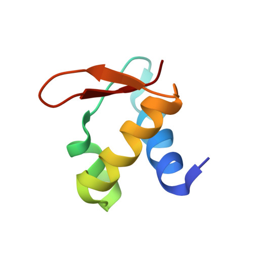
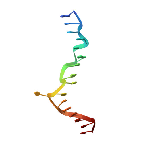
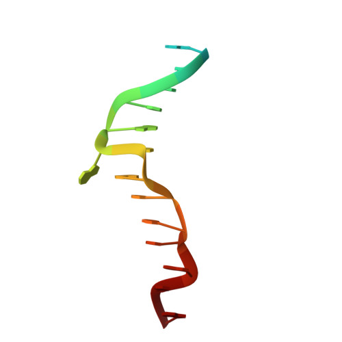
Sequence preference and structural heterogeneity of BZ junctions.

Kim, D.Hur, J.Han, J.H.Ha, S.C.Shin, D.Lee, S.Park, S.Sugiyama, H.Kim, K.K.

(2018) Nucleic Acids Res 46: 10504-10513

  • DOI: https://doi.org/10.1093/nar/gky784
  • Primary Citation Related Structures: 
    5ZU1, 5ZUO, 5ZUP

  • PubMed Abstract: 

    BZ junctions, which connect B-DNA to Z-DNA, are necessary for local transformation of B-DNA to Z-DNA in the genome. However, the limited information on the junction-forming sequences and junction structures has led to a lack of understanding of the structural diversity and sequence preferences of BZ junctions. We determined three crystal structures of BZ junctions with diverse sequences followed by spectroscopic validation of DNA conformation. The structural features of the BZ junctions were well conserved regardless of sequences via the continuous base stacking through B-to-Z DNA with A-T base extrusion. However, the sequence-dependent structural heterogeneity of the junctions was also observed in base step parameters that are correlated with steric constraints imposed during Z-DNA formation. Further, circular dichroism and fluorescence-based analysis of BZ junctions revealed that a base extrusion was only found at the A-T base pair present next to a stable dinucleotide Z-DNA unit. Our findings suggest that Z-DNA formation in the genome is influenced by the sequence preference for BZ junctions.


  • Organizational Affiliation
    • Department of Molecular Cell Biology, Institute for Antimicrobial Research and Therapeutics, Sungkyunkwan University School of Medicine, Suwon 16419, Korea.

Macromolecule Content 

  • Total Structure Weight: 38.25 kDa 
  • Atom Count: 2,372 
  • Modeled Residue Count: 262 
  • Deposited Residue Count: 286 
  • Unique protein chains: 1
  • Unique nucleic acid chains: 2

Macromolecules


Find similar proteins by:|  3D Structure
Entity ID: 1
MoleculeChains  Sequence LengthOrganismDetailsImage
Double-stranded RNA-specific adenosine deaminase
A, B, C, D
63Homo sapiensMutation(s): 0 
Gene Names: ADARADAR1DSRADG1P1IFI4
EC: 3.5.4.37
UniProt & NIH Common Fund Data Resources
Find proteins for P55265 (Homo sapiens)
Explore P55265 
Go to UniProtKB:  P55265
PHAROS:  P55265
GTEx:  ENSG00000160710 
Entity Groups
Sequence Clusters30% Identity50% Identity70% Identity90% Identity95% Identity100% Identity
UniProt GroupP55265
Sequence Annotations
Expand
Reference Sequence
Find similar nucleic acids by:  Sequence
Entity ID: 2
MoleculeChains LengthOrganismImage
DNA (5'-D(*GP*TP*CP*GP*CP*GP*CP*GP*CP*CP*TP*TP*AP*AP*AP*CP*C)-3')17synthetic construct
Sequence Annotations
Expand
Reference Sequence
Find similar nucleic acids by:  Sequence
Entity ID: 3
MoleculeChains LengthOrganismImage
DNA (5'-D(*AP*CP*GP*GP*TP*TP*TP*AP*AP*GP*GP*CP*GP*CP*GP*CP*G)-3')17synthetic construct
Sequence Annotations
Expand
Reference Sequence

Experimental Data & Validation

Experimental Data

  • Method: X-RAY DIFFRACTION
  • Resolution: 3.01 Å
  • R-Value Free:  0.280 (Depositor), 0.279 (DCC) 
  • R-Value Work:  0.233 (Depositor), 0.232 (DCC) 
  • R-Value Observed: 0.238 (Depositor) 
Space Group: P 61
Unit Cell:
Length ( Å )Angle ( ˚ )
a = 108.92α = 90
b = 108.92β = 90
c = 62.004γ = 120
Software Package:
Software NamePurpose
HKL-2000data scaling
PHENIXrefinement
PDB_EXTRACTdata extraction
HKL-2000data reduction
MOLREPphasing

Structure Validation

View Full Validation Report


Entry History 

& Funding Information

Deposition Data


Funding OrganizationLocationGrant Number
Korea, Republic OfSSTF-BA1301-01

Revision History  (Full details and data files)

  • Version 1.0: 2018-08-29
    Type: Initial release
  • Version 1.1: 2018-09-19
    Changes: Data collection, Database references
  • Version 1.2: 2018-11-21
    Changes: Data collection, Database references
  • Version 1.3: 2023-11-22
    Changes: Data collection, Database references, Refinement description