5ZR2 | pdb_00005zr2

Crystal Structure of Phosphoserine Phosphatase Mutant (H9A) from Entamoeba histolytica in complex with Phosphoserine


Experimental Data Snapshot

  • Method: X-RAY DIFFRACTION
  • Resolution: 2.95 Å
  • R-Value Free: 
    0.250 (Depositor), 0.245 (DCC) 
  • R-Value Work: 
    0.207 (Depositor), 0.208 (DCC) 
  • R-Value Observed: 
    0.209 (Depositor) 

Starting Model: experimental
View more details

wwPDB Validation 3D Report Full Report

Validation slider image for 5ZR2

Ligand Structure Quality Assessment 


This is version 1.2 of the entry. See complete history

Literature

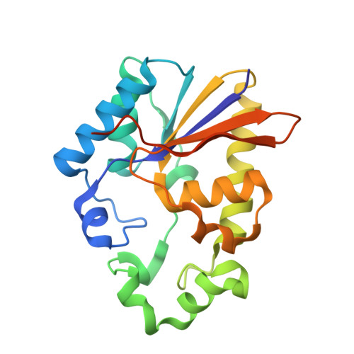
Structural and functional characterisation of phosphoserine phosphatase, that plays critical role in the oxidative stress response in the parasite Entamoeba histolytica.

Kumari, P.Babuta, M.Bhattacharya, A.Gourinath, S.

(2019) J Struct Biol 206: 254-266

  • DOI: https://doi.org/10.1016/j.jsb.2019.03.012
  • Primary Citation Related Structures: 
    5ZKK, 5ZR2

  • PubMed Abstract: 

    Amoebiasis is a common parasitic infection in the developing world and is caused by the protist Entameoba histolytica. The proliferation of E. histolytica and its ability to invade epithelial tissues have been shown in several studies to be greatly decreased during oxidative stress. It is therefore not surprising that this amoeba has evolved several mechanisms to evade oxidative stress. Cysteine is thought to be one of the crucial molecules that help in redox defence, and a de novo cysteine biosynthetic pathway involving serine as one of the substrates has been partially elucidated in E. histolytica. Though most of the enzymes of this pathway in E. histolytica have been characterized, phosphoserine phosphatase (EhPSP), a key regulatory enzyme of the serine biosynthetic pathway, has not yet even been identified. In the current work, we identified and characterized EhPSP using various molecular, structural and functional approaches. The crystal structures of native and substrate-bound EhPSP were determined and showed the residues that play a crucial role in its phosphatase activity and substrate binding. Structural and biochemical studies indicated that EhPSP belongs to the histidine phosphatase superfamily. EhPSP-overexpressing amoebic cells were found to be more tolerant to oxidative stress. However, protection during oxidative stress was not seen when a functionally defective mutant was overexpressed. Our results clearly showed that E. histolytica has a functional PSP and that this protein participates in protecting the organism against oxidative stress.


  • Organizational Affiliation
    • School of Life Sciences, Jawaharlal Nehru University, New Delhi, India.

Macromolecule Content 

  • Total Structure Weight: 96.05 kDa 
  • Atom Count: 6,177 
  • Modeled Residue Count: 789 
  • Deposited Residue Count: 844 
  • Unique protein chains: 1

Macromolecules

Find similar proteins by:|  3D Structure
Entity ID: 1
MoleculeChains  Sequence LengthOrganismDetailsImage
Phosphoglycerate mutase family protein, putative
A, B, C, D
211Entamoeba histolytica HM-1:IMSS-AMutation(s): 1 
Gene Names: EHI7A_041450
UniProt
Find proteins for N9V397 (Entamoeba histolytica HM-1:IMSS-A)
Explore N9V397 
Go to UniProtKB:  N9V397
Entity Groups
Sequence Clusters30% Identity50% Identity70% Identity90% Identity95% Identity100% Identity
UniProt GroupN9V397
Sequence Annotations
Expand
Reference Sequence

Small Molecules

Ligands 4 Unique
IDChains Name / Formula / InChI Key2D Diagram3D Interactions
PG4

Query on PG4


Download:Ideal Coordinates CCD File
G [auth A]TETRAETHYLENE GLYCOL
C8 H18 O5
UWHCKJMYHZGTIT-UHFFFAOYSA-N
SEP

Query on SEP


Download:Ideal Coordinates CCD File
E [auth A],
H [auth B],
J [auth C],
L [auth D]
PHOSPHOSERINE
C3 H8 N O6 P
BZQFBWGGLXLEPQ-REOHCLBHSA-N
PEG

Query on PEG


Download:Ideal Coordinates CCD File
I [auth B]DI(HYDROXYETHYL)ETHER
C4 H10 O3
MTHSVFCYNBDYFN-UHFFFAOYSA-N
SO4

Query on SO4


Download:Ideal Coordinates CCD File
F [auth A],
K [auth C]
SULFATE ION
O4 S
QAOWNCQODCNURD-UHFFFAOYSA-L

Experimental Data & Validation

Experimental Data

  • Method: X-RAY DIFFRACTION
  • Resolution: 2.95 Å
  • R-Value Free:  0.250 (Depositor), 0.245 (DCC) 
  • R-Value Work:  0.207 (Depositor), 0.208 (DCC) 
  • R-Value Observed: 0.209 (Depositor) 
Space Group: C 2 2 21
Unit Cell:
Length ( Å )Angle ( ˚ )
a = 95.107α = 90
b = 111.737β = 90
c = 178.246γ = 90
Software Package:
Software NamePurpose
REFMACrefinement
HKL-2000data reduction
HKL-2000data scaling
PHASERphasing

Structure Validation

View Full Validation Report


Ligand Structure Quality Assessment 


Entry History 

Deposition Data

Revision History  (Full details and data files)

  • Version 1.0: 2019-04-24
    Type: Initial release
  • Version 1.1: 2019-05-15
    Changes: Data collection, Database references
  • Version 1.2: 2023-11-22
    Changes: Data collection, Database references, Refinement description