


Crystal structure of Oryza sativa hexokinase 6
Matsudaira, K., Mochizuki, S., Yoshida, H., Kamitori, S., Akimitsu, K.To be published.
Experimental Data Snapshot
Starting Model: experimental
View more details
Entity ID: 1 | |||||
|---|---|---|---|---|---|
| Molecule | Chains | Sequence Length | Organism | Details | Image |
| Hexokinase-6 | 473 | Oryza sativa Japonica Group | Mutation(s): 0 Gene Names: HXK6, HXK2, Os01g0742500, LOC_Os01g53930, P0439E07.19 EC: 2.7.1.1 | 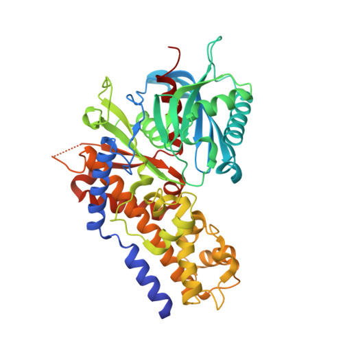 | |
UniProt | |||||
Entity Groups | |||||
| Sequence Clusters | 30% Identity50% Identity70% Identity90% Identity95% Identity100% Identity | ||||
| UniProt Group | Q8LQ68 | ||||
Sequence AnnotationsExpand | |||||
Reference Sequence | |||||
| Ligands 3 Unique | |||||
|---|---|---|---|---|---|
| ID | Chains | Name / Formula / InChI Key | 2D Diagram | 3D Interactions | |
| ANP (Subject of Investigation/LOI) Download:Ideal Coordinates CCD File | D [auth A], G [auth B], J [auth C] | PHOSPHOAMINOPHOSPHONIC ACID-ADENYLATE ESTER C10 H17 N6 O12 P3 PVKSNHVPLWYQGJ-KQYNXXCUSA-N |  | ||
| BGC (Subject of Investigation/LOI) Download:Ideal Coordinates CCD File | F [auth A], I [auth B], L [auth C] | beta-D-glucopyranose C6 H12 O6 WQZGKKKJIJFFOK-VFUOTHLCSA-N |  | ||
| MG (Subject of Investigation/LOI) Download:Ideal Coordinates CCD File | E [auth A], H [auth B], K [auth C] | MAGNESIUM ION Mg JLVVSXFLKOJNIY-UHFFFAOYSA-N |  | ||
| Length ( Å ) | Angle ( ˚ ) |
|---|---|
| a = 131.6 | α = 90 |
| b = 131.6 | β = 90 |
| c = 188.92 | γ = 120 |
| Software Name | Purpose |
|---|---|
| REFMAC | refinement |
| XDS | data reduction |
| XDS | data scaling |
| MOLREP | phasing |


