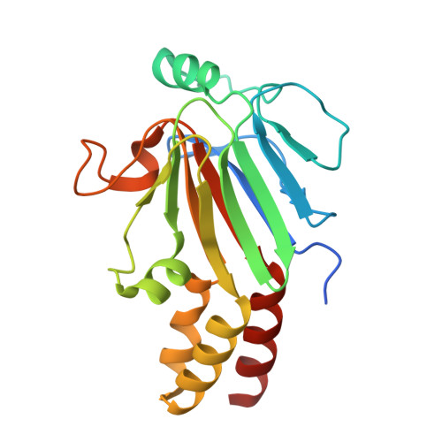
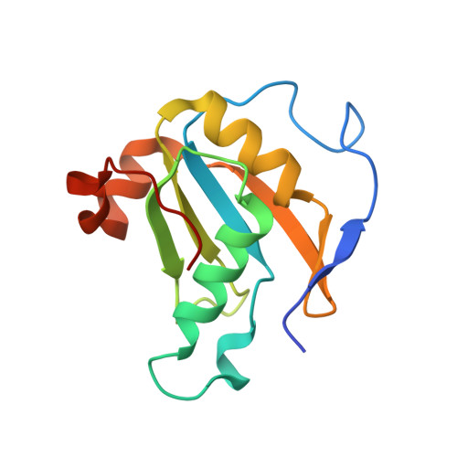
5ZOJ

Crystal structure of human SMAD2-MAN1 complex


Experimental Data Snapshot

  • Method: X-RAY DIFFRACTION
  • Resolution: 2.79 Å
  • R-Value Free: 0.269 
  • R-Value Work: 0.242 
  • R-Value Observed: 0.243 

wwPDB Validation   3D Report Full Report


This is version 1.3 of the entry. See complete history


Literature

Structural basis for receptor-regulated SMAD recognition by MAN1

Miyazono, K.I.Ohno, Y.Wada, H.Ito, T.Fukatsu, Y.Kurisaki, A.Asashima, M.Tanokura, M.

(2018) Nucleic Acids Res 46: 12139-12153

  • DOI: https://doi.org/10.1093/nar/gky925
  • Primary Citation of Related Structures:  
    5ZOJ, 5ZOK

  • PubMed Abstract: 

    Receptor-regulated SMAD (R-SMAD: SMAD1, SMAD2, SMAD3, SMAD5 and SMAD8) proteins are key transcription factors of the transforming growth factor-β (TGF-β) superfamily of cytokines. MAN1, an integral protein of the inner nuclear membrane, is a SMAD cofactor that terminates TGF-β superfamily signals. Heterozygous loss-of-function mutations in MAN1 result in osteopoikilosis, Buschke-Ollendorff syndrome and melorheostosis. MAN1 interacts with MAD homology 2 (MH2) domains of R-SMAD proteins using its C-terminal U2AF homology motif (UHM) domain and UHM ligand motif (ULM) and facilitates R-SMAD dephosphorylation. Here, we report the structural basis for R-SMAD recognition by MAN1. The SMAD2-MAN1 and SMAD1-MAN1 complex structures show that an intramolecular UHM-ULM interaction of MAN1 forms a hydrophobic surface that interacts with a hydrophobic surface among the H2 helix, the strands β8 and β9, and the L3 loop of the MH2 domains of R-SMAD proteins. The complex structures also show the mechanism by which SMAD cofactors distinguish R-SMAD proteins that possess a highly conserved molecular surface.


  • Organizational Affiliation

    Department of Applied Biological Chemistry, Graduate School of Agricultural and Life Sciences, The University of Tokyo, Tokyo 113-8657, Japan.


Macromolecules
Find similar proteins by:  (by identity cutoff)  |  3D Structure
Entity ID: 1
MoleculeChains Sequence LengthOrganismDetailsImage
Mothers against decapentaplegic homolog 2
A, B, C
200Homo sapiensMutation(s): 0 
Gene Names: SMAD2
UniProt & NIH Common Fund Data Resources
Find proteins for Q15796 (Homo sapiens)
Explore Q15796 
Go to UniProtKB:  Q15796
PHAROS:  Q15796
GTEx:  ENSG00000175387 
Entity Groups  
Sequence Clusters30% Identity50% Identity70% Identity90% Identity95% Identity100% Identity
UniProt GroupQ15796
Sequence Annotations
Expand
  • Reference Sequence
Find similar proteins by:  (by identity cutoff)  |  3D Structure
Entity ID: 2
MoleculeChains Sequence LengthOrganismDetailsImage
Inner nuclear membrane protein Man1
D, E
132Homo sapiensMutation(s): 0 
Gene Names: MAN1
UniProt & NIH Common Fund Data Resources
Find proteins for Q9Y2U8 (Homo sapiens)
Explore Q9Y2U8 
Go to UniProtKB:  Q9Y2U8
PHAROS:  Q9Y2U8
GTEx:  ENSG00000174106 
Entity Groups  
Sequence Clusters30% Identity50% Identity70% Identity90% Identity95% Identity100% Identity
UniProt GroupQ9Y2U8
Sequence Annotations
Expand
  • Reference Sequence
Experimental Data & Validation

Experimental Data

  • Method: X-RAY DIFFRACTION
  • Resolution: 2.79 Å
  • R-Value Free: 0.269 
  • R-Value Work: 0.242 
  • R-Value Observed: 0.243 
  • Space Group: P 65
Unit Cell:
Length ( Å )Angle ( ˚ )
a = 176.813α = 90
b = 176.813β = 90
c = 73.854γ = 120
Software Package:
Software NamePurpose
Aimlessdata scaling
PHENIXrefinement
PDB_EXTRACTdata extraction
XDSdata reduction
MOLREPphasing

Structure Validation

View Full Validation Report


Entry History & Funding Information

Deposition Data


Funding OrganizationLocationGrant Number
Japan Society for the Promotion of ScienceJapan15K14708
Japan Society for the Promotion of ScienceJapan17K19581
Japan Society for the Promotion of ScienceJapan23228003

Revision History  (Full details and data files)

  • Version 1.0: 2018-10-10
    Type: Initial release
  • Version 1.1: 2018-11-14
    Changes: Data collection, Database references, Structure summary
  • Version 1.2: 2018-12-26
    Changes: Data collection, Database references
  • Version 1.3: 2024-03-27
    Changes: Data collection, Database references, Refinement description