5ZO5 | pdb_00005zo5

active state of the nuclease


Experimental Data Snapshot

  • Method: X-RAY DIFFRACTION
  • Resolution: 2.30 Å
  • R-Value Free: 
    0.225 (Depositor), 0.229 (DCC) 
  • R-Value Work: 
    0.186 (Depositor), 0.188 (DCC) 
  • R-Value Observed: 
    0.189 (Depositor) 

wwPDB Validation 3D Report Full Report

Validation slider image for 5ZO5

This is version 1.1 of the entry. See complete history

Literature

NrnC, an RNase D-Like Protein FromAgrobacterium, Is a Novel Octameric Nuclease That Specifically Degrades dsDNA but Leaves dsRNA Intact.

Yuan, Z.Gao, F.Yin, K.Gu, L.

(2018) Front Microbiol 9: 3230-3230

  • DOI: https://doi.org/10.3389/fmicb.2018.03230
  • Primary Citation Related Structures: 
    5ZO3, 5ZO4, 5ZO5

  • PubMed Abstract: 

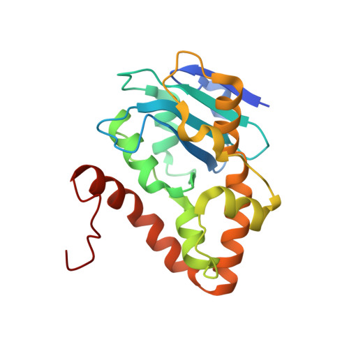
    NrnC from Agrobacterium tumefaciens (At_NrnC, UniProt accession number A9CG28) is a nuclease containing a single DEDDy domain. Here, we determined the structures of both the apo and metal-ion-bound forms of At_NrnC. Although the overall structure of the At_NrnC protomer is similar to that of the RNase D exonuclease domain, nuclease assays unexpectedly revealed that At_NrnC possesses remarkably different substrate specificity. In contrast to RNase D, which degrades both single-stranded RNA (ssRNA) and double-stranded RNA (dsRNA), At_NrnC hydrolyses ssRNA, single-stranded DNA (ssDNA), and double-stranded DNA (dsDNA) with high efficiency but does not degrade dsRNA. Crystal packing analysis and biochemical data indicated that At_NrnC forms an octameric hollow cylindrical structure that allows ssRNA, ssDNA, and dsDNA, but not dsRNA, to enter the central tunnel where the multiple active sites perform hydrolysis. This novel structural feature confers a high processivity and is responsible for the preference of At_NrnC for longer dsDNA substrates.


  • Organizational Affiliation
    • State Key Laboratory of Microbial Technology, Shandong University, Jinan, China.

Macromolecule Content 

  • Total Structure Weight: 47.6 kDa 
  • Atom Count: 3,514 
  • Modeled Residue Count: 411 
  • Deposited Residue Count: 424 
  • Unique protein chains: 1

Macromolecules

Find similar proteins by:|  3D Structure
Entity ID: 1
MoleculeChains  Sequence LengthOrganismDetailsImage
Putative 3'-5' exonuclease family protein
A, B
212Agrobacterium fabrum str. J-07Mutation(s): 0 
Gene Names: AGR8A_Lc10769
EC: 3.1.13

Small Molecules

Ligands 2 Unique
IDChains Name / Formula / InChI Key2D Diagram3D Interactions
SO4

Query on SO4


Download:Ideal Coordinates CCD File
E [auth A],
F [auth A],
G [auth A],
J [auth B],
K [auth B]
SULFATE ION
O4 S
QAOWNCQODCNURD-UHFFFAOYSA-L
MN

Query on MN


Download:Ideal Coordinates CCD File
C [auth A],
D [auth A],
H [auth B],
I [auth B]
MANGANESE (II) ION
Mn
WAEMQWOKJMHJLA-UHFFFAOYSA-N

Experimental Data & Validation

Experimental Data

  • Method: X-RAY DIFFRACTION
  • Resolution: 2.30 Å
  • R-Value Free:  0.225 (Depositor), 0.229 (DCC) 
  • R-Value Work:  0.186 (Depositor), 0.188 (DCC) 
  • R-Value Observed: 0.189 (Depositor) 
Space Group: I 4 2 2
Unit Cell:
Length ( Å )Angle ( ˚ )
a = 121.743α = 90
b = 121.743β = 90
c = 149.971γ = 90
Software Package:
Software NamePurpose
PHENIXrefinement
HKL-2000data reduction
HKL-2000data scaling
PHENIXphasing

Structure Validation

View Full Validation Report


Entry History 

Deposition Data

Revision History  (Full details and data files)

  • Version 1.0: 2019-04-10
    Type: Initial release
  • Version 1.1: 2024-03-27
    Changes: Data collection, Database references, Derived calculations