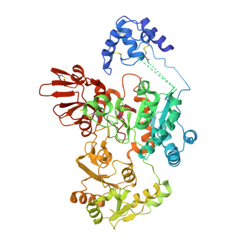
5ZIB

Crystal structure of human GnT-V luminal domain in apo form


Experimental Data Snapshot

  • Method: X-RAY DIFFRACTION
  • Resolution: 1.90 Å
  • R-Value Free: 0.224 
  • R-Value Work: 0.189 
  • R-Value Observed: 0.191 

wwPDB Validation   3D Report Full Report


Ligand Structure Quality Assessment 


This is version 1.3 of the entry. See complete history


Literature

Structure and mechanism of cancer-associated N-acetylglucosaminyltransferase-V.

Nagae, M.Kizuka, Y.Mihara, E.Kitago, Y.Hanashima, S.Ito, Y.Takagi, J.Taniguchi, N.Yamaguchi, Y.

(2018) Nat Commun 9: 3380-3380

  • DOI: https://doi.org/10.1038/s41467-018-05931-w
  • Primary Citation of Related Structures:  
    5ZIB, 5ZIC

  • PubMed Abstract: 

    N-acetylglucosaminyltransferase-V (GnT-V) alters the structure of specific N-glycans by modifying α1-6-linked mannose with a β1-6-linked N-acetylglucosamine branch. β1-6 branch formation on cell surface receptors accelerates cancer metastasis, making GnT-V a promising target for drug development. However, the molecular basis of GnT-V's catalytic mechanism and substrate specificity are not fully understood. Here, we report crystal structures of human GnT-V luminal domain with a substrate analog. GnT-V luminal domain is composed of a GT-B fold and two accessary domains. Interestingly, two aromatic rings sandwich the α1-6 branch of the acceptor N-glycan and restrain the global conformation, partly explaining the fine branch specificity of GnT-V. In addition, interaction of the substrate N-glycoprotein with GnT-V likely contributes to protein-selective and site-specific glycan modification. In summary, the acceptor-GnT-V complex structure suggests a catalytic mechanism, explains the previously observed inhibition of GnT-V by branching enzyme GnT-III, and provides a basis for the rational design of drugs targeting N-glycan branching.


  • Organizational Affiliation

    Structural Glycobiology Team, Glycobiology Research Group, Global Research Cluster, RIKEN, 2-1 Hirosawa, Wako, Saitama, 351-0198, Japan. mnagae@mol.f.u-tokyo.ac.jp.


Macromolecules
Find similar proteins by:  (by identity cutoff)  |  3D Structure
Entity ID: 1
MoleculeChains Sequence LengthOrganismDetailsImage
Alpha-1,6-mannosylglycoprotein 6-beta-N-acetylglucosaminyltransferase A626Homo sapiensMutation(s): 0 
Gene Names: MGAT5GGNT5
EC: 2.4.1.155
UniProt & NIH Common Fund Data Resources
Find proteins for Q09328 (Homo sapiens)
Explore Q09328 
Go to UniProtKB:  Q09328
PHAROS:  Q09328
GTEx:  ENSG00000152127 
Entity Groups  
Sequence Clusters30% Identity50% Identity70% Identity90% Identity95% Identity100% Identity
UniProt GroupQ09328
Glycosylation
Glycosylation Sites: 1Go to GlyGen: Q09328-1
Sequence Annotations
Expand
  • Reference Sequence
Small Molecules
Ligands 1 Unique
IDChains Name / Formula / InChI Key2D Diagram3D Interactions
NAG
Query on NAG

Download Ideal Coordinates CCD File 
B [auth A]2-acetamido-2-deoxy-beta-D-glucopyranose
C8 H15 N O6
OVRNDRQMDRJTHS-FMDGEEDCSA-N
Experimental Data & Validation

Experimental Data

  • Method: X-RAY DIFFRACTION
  • Resolution: 1.90 Å
  • R-Value Free: 0.224 
  • R-Value Work: 0.189 
  • R-Value Observed: 0.191 
  • Space Group: P 65 2 2
Unit Cell:
Length ( Å )Angle ( ˚ )
a = 97.671α = 90
b = 97.671β = 90
c = 268.856γ = 120
Software Package:
Software NamePurpose
PHENIXrefinement
XDSdata reduction
Aimlessdata scaling
AutoSolphasing

Structure Validation

View Full Validation Report


Ligand Structure Quality Assessment 


Entry History & Funding Information

Deposition Data


Funding OrganizationLocationGrant Number
MEXTJapan17K07303

Revision History  (Full details and data files)

  • Version 1.0: 2018-08-01
    Type: Initial release
  • Version 1.1: 2018-09-05
    Changes: Data collection, Database references
  • Version 1.2: 2020-07-29
    Type: Remediation
    Reason: Carbohydrate remediation
    Changes: Data collection, Derived calculations, Structure summary
  • Version 1.3: 2024-10-09
    Changes: Data collection, Database references, Structure summary