5ZDS | pdb_00005zds

Crystal structure of the second PDZ domain of Frmpd2


Experimental Data Snapshot

  • Method: X-RAY DIFFRACTION
  • Resolution: 1.80 Å
  • R-Value Free: 
    0.235 (Depositor), 0.236 (DCC) 
  • R-Value Work: 
    0.198 (Depositor), 0.200 (DCC) 
  • R-Value Observed: 
    0.202 (Depositor) 

Starting Model: experimental
View more details

wwPDB Validation 3D Report Full Report

Validation slider image for 5ZDS

This is version 1.3 of the entry. See complete history

Literature

The second PDZ domain of scaffold protein Frmpd2 binds to GluN2A of NMDA receptors.

Lu, X.Zhang, Q.Wang, T.

(2019) Biochem Biophys Res Commun 516: 63-67

  • DOI: https://doi.org/10.1016/j.bbrc.2019.05.087
  • Primary Citation Related Structures: 
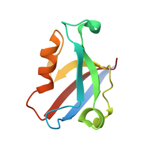
    5ZDS

  • PubMed Abstract: 

    The scaffold proteins Frmpd2 is localized at the basolateral membranes of polarized epithelial cells and associated with tight junction formation. In this report, we found that the Frmpd2 is specifically expressed at postsynaptic membrane. By using of co-immunoprecipitation and GST pull-down, Frmpd2 was reported to interact with postsynaptic excitatory N-methyl-d-aspartic acid (NMDA) receptors in vivo and in vitro. In addition, we demonstrated that the second PDZ (PDZ2) domain but not the first or third PDZ domain of Frmpd2 binds to the C-terminus of GluN2A and GluN2B, two subunits of NMDA receptors. By surface plasmon resonance, the affinity of Frmpd2-isolated PDZ2 to GluN2A and GluN2B was identified, which indicates that the interaction of Frmpd2 to GluN2A subunit is more strongly than that to GluN2B subunit. The crystal structure of the PDZ2 domain of the mouse homologue of Frmpd2 was further solved. Some amino acid residues of the PDZ2 structure are supposed to associate with the GluN2A binding. Our study suggests that the scaffold protein Frmpd2 is probably involved in synaptic NMDA receptors-mediated neural excitatory and neurotoxicity in a PDZ2 domain-dependent manner.


  • Organizational Affiliation
    • Department of Neurology, Chongqing Key Laboratory of Neurology, The First Affiliated Hospital of Chongqing Medical University, 1 Youyi Road, Chongqing, 400016, China. Electronic address: lxi2646@163.com.

Macromolecule Content 

  • Total Structure Weight: 10 kDa 
  • Atom Count: 810 
  • Modeled Residue Count: 88 
  • Deposited Residue Count: 96 
  • Unique protein chains: 1

Macromolecules

Find similar proteins by:|  3D Structure
Entity ID: 1
MoleculeChains  Sequence LengthOrganismDetailsImage
FERM and PDZ domain-containing 296Mus musculusMutation(s): 0 
Gene Names: Frmpd2Gm626
UniProt
Find proteins for A0A140LI67 (Mus musculus)
Explore A0A140LI67 
Go to UniProtKB:  A0A140LI67
Entity Groups
Sequence Clusters30% Identity50% Identity70% Identity90% Identity95% Identity100% Identity
UniProt GroupA0A140LI67
Sequence Annotations
Expand
Reference Sequence

Experimental Data & Validation

Experimental Data

  • Method: X-RAY DIFFRACTION
  • Resolution: 1.80 Å
  • R-Value Free:  0.235 (Depositor), 0.236 (DCC) 
  • R-Value Work:  0.198 (Depositor), 0.200 (DCC) 
  • R-Value Observed: 0.202 (Depositor) 
Space Group: P 21 21 2
Unit Cell:
Length ( Å )Angle ( ˚ )
a = 48.176α = 90
b = 56.842β = 90
c = 32.2γ = 90
Software Package:
Software NamePurpose
PHENIXrefinement
HKL-3000data reduction
HKL-3000data scaling
PHENIXphasing

Structure Validation

View Full Validation Report


Entry History 

Deposition Data

Revision History  (Full details and data files)

  • Version 1.0: 2019-02-27
    Type: Initial release
  • Version 1.1: 2023-11-22
    Changes: Data collection, Database references, Refinement description
  • Version 1.2: 2024-04-03
    Changes: Database references
  • Version 1.3: 2024-11-20
    Changes: Structure summary