


Crystal structure of sugar-binding protein YesO in complex with rhamnogalacturonan trisaccharide
Sugiura, H., Oiki, S., Mikami, B., Murata, K., Hashimoto, W.To be published.
Experimental Data Snapshot
Starting Model: experimental
View more details
wwPDB Validation 3D Report Full Report

Entity ID: 1 | |||||
|---|---|---|---|---|---|
| Molecule | Chains | Sequence Length | Organism | Details | Image |
| Putative ABC transporter substrate-binding protein YesO | 427 | Bacillus subtilis subsp. subtilis str. 168 | Mutation(s): 0 Gene Names: yesO, BSU06970 | 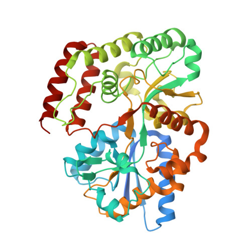 | |
UniProt | |||||
Entity Groups | |||||
| Sequence Clusters | 30% Identity50% Identity70% Identity90% Identity95% Identity100% Identity | ||||
| UniProt Group | O31518 | ||||
Sequence AnnotationsExpand | |||||
Reference Sequence | |||||
| Length ( Å ) | Angle ( ˚ ) |
|---|---|
| a = 42.23 | α = 90 |
| b = 78.592 | β = 102.04 |
| c = 54.075 | γ = 90 |
| Software Name | Purpose |
|---|---|
| PHENIX | refinement |
| HKL-2000 | data reduction |
| HKL-2000 | data scaling |
| MOLREP | phasing |
| Funding Organization | Location | Grant Number |
|---|---|---|
| Fuji Foundation for Protein Research | Japan | -- |


