5YVX

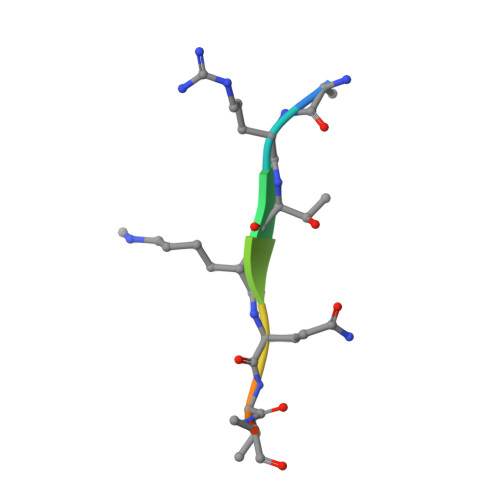
Crystal structure of SDG8 CW domain in complex with H3K4me1 peptide


Experimental Data Snapshot

  • Method: X-RAY DIFFRACTION
  • Resolution: 1.59 Å
  • R-Value Free: 0.184 
  • R-Value Work: 0.168 
  • R-Value Observed: 0.169 

wwPDB Validation   3D Report Full Report


This is version 1.1 of the entry. See complete history


Literature

Uncovering the mechanistic basis for specific recognition of monomethylated H3K4 by the CW domain ofArabidopsishistone methyltransferase SDG8.

Liu, Y.Huang, Y.

(2018) J Biol Chem 293: 6470-6481

  • DOI: https://doi.org/10.1074/jbc.RA117.001390
  • Primary Citation of Related Structures:  
    5YVX

  • PubMed Abstract: 

    Chromatin consists of DNA and histones, and specific histone modifications that determine chromatin structure and activity are regulated by three types of proteins, called writer, reader, and eraser. Histone reader proteins from vertebrates, vertebrate-infecting parasites, and higher plants possess a CW domain, which has been reported to read histone H3 lysine 4 (H3K4). The CW domain of Arabidopsis SDG8 (also called ASHH2), a histone H3 lysine 36 methyltransferase, preferentially binds monomethylated H3K4 (H3K4me1), unlike the mammalian CW domain protein, which binds trimethylated H3K4 (H3K4me3). However, the molecular basis of the selective binding by the CW domain of SDG8 (SDG8-CW) remains unclear. Here, we solved the 1.6-Å-resolution structure of SDG8-CW in complex with H3K4me1, which revealed that residues in the C-terminal α-helix of SDG8-CW determine binding specificity for low methylation levels at H3K4. Moreover, substitutions of key residues, specifically Ile-915 and Asn-916, converted SDG8-CW binding preference from H3K4me1 to H3K4me3. Sequence alignment and mutagenesis studies revealed that the CW domain of SDG725, the homolog of SDG8 in rice, shares the same binding preference with SDG8-CW, indicating that preference for low methylated H3K4 by the CW domain of ASHH2 homologs is conserved among higher-order plants. Our findings provide first structural insights into the molecular basis for specific recognition of monomethylated H3K4 by the H3K4me1 reader protein SDG8 from Arabidopsis .


  • Organizational Affiliation

    From the State Key Laboratory of Molecular Biology, National Center for Protein Science Shanghai, Shanghai Science Research Center, Shanghai Key Laboratory of Molecular Andrology, CAS Center for Excellence in Molecular Cell Science, Shanghai Institute of Biochemistry and Cell Biology, Chinese Academy of Sciences, University of Chinese Academy of Sciences, Shanghai 201210, China.


Macromolecules
Find similar proteins by:  (by identity cutoff)  |  3D Structure
Entity ID: 1
MoleculeChains Sequence LengthOrganismDetailsImage
Histone-lysine N-methyltransferase ASHH260Arabidopsis thalianaMutation(s): 1 
Gene Names: ASHH2EFSLAZ2SDG8SET8At1g77300T14N5.15
EC: 2.1.1.43 (PDB Primary Data), 2.1.1 (UniProt)
UniProt
Find proteins for Q2LAE1 (Arabidopsis thaliana)
Explore Q2LAE1 
Go to UniProtKB:  Q2LAE1
Entity Groups  
Sequence Clusters30% Identity50% Identity70% Identity90% Identity95% Identity100% Identity
UniProt GroupQ2LAE1
Sequence Annotations
Expand
  • Reference Sequence

Find similar proteins by:  Sequence   |   3D Structure  

Entity ID: 2
MoleculeChains Sequence LengthOrganismDetailsImage
H3K4me1B [auth C]9Arabidopsis thalianaMutation(s): 0 
UniProt
Find proteins for P59226 (Arabidopsis thaliana)
Explore P59226 
Go to UniProtKB:  P59226
Entity Groups  
UniProt GroupP59226
Sequence Annotations
Expand
  • Reference Sequence
Small Molecules
Ligands 1 Unique
IDChains Name / Formula / InChI Key2D Diagram3D Interactions
ZN
Query on ZN

Download Ideal Coordinates CCD File 
C [auth A]ZINC ION
Zn
PTFCDOFLOPIGGS-UHFFFAOYSA-N
Modified Residues  1 Unique
IDChains TypeFormula2D DiagramParent
MLZ
Query on MLZ
B [auth C]L-PEPTIDE LINKINGC7 H16 N2 O2LYS
Experimental Data & Validation

Experimental Data

  • Method: X-RAY DIFFRACTION
  • Resolution: 1.59 Å
  • R-Value Free: 0.184 
  • R-Value Work: 0.168 
  • R-Value Observed: 0.169 
  • Space Group: P 63
Unit Cell:
Length ( Å )Angle ( ˚ )
a = 66.421α = 90
b = 66.421β = 90
c = 29.241γ = 120
Software Package:
Software NamePurpose
PHENIXrefinement
HKL-3000data reduction
HKL-3000data scaling
PHENIXphasing

Structure Validation

View Full Validation Report


Entry History 

Deposition Data

Revision History  (Full details and data files)

  • Version 1.0: 2018-03-14
    Type: Initial release
  • Version 1.1: 2018-05-09
    Changes: Data collection, Database references