


Crystal structure of E.coli aminopeptidase N in complex with Puromycin aminonucleoside
Marapaka, A.K., Ganji, R.J., Reddi, R., Addlagatta, A.To be published.
Experimental Data Snapshot
Starting Model: experimental
View more details
Entity ID: 1 | |||||
|---|---|---|---|---|---|
| Molecule | Chains | Sequence Length | Organism | Details | Image |
| Aminopeptidase N | 891 | Escherichia coli K-12 | Mutation(s): 0 Gene Names: pepN, b0932, JW0915 EC: 3.4.11.2 | 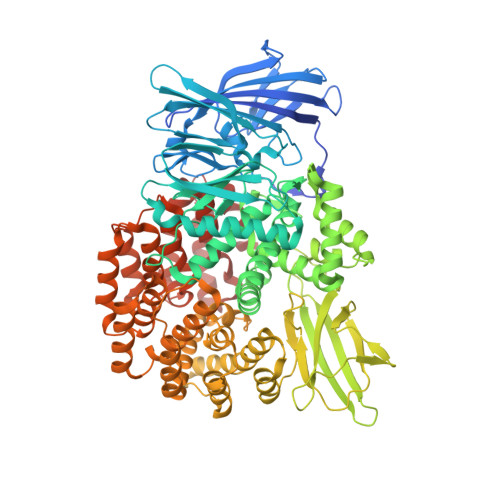 | |
UniProt | |||||
Entity Groups | |||||
| Sequence Clusters | 30% Identity50% Identity70% Identity90% Identity95% Identity100% Identity | ||||
| UniProt Group | P04825 | ||||
Sequence AnnotationsExpand | |||||
Reference Sequence | |||||
| Ligands 4 Unique | |||||
|---|---|---|---|---|---|
| ID | Chains | Name / Formula / InChI Key | 2D Diagram | 3D Interactions | |
| GMC Download:Ideal Coordinates CCD File | C [auth A] | (2R,3R,4S,5S)-4-AMINO-2-[6-(DIMETHYLAMINO)-9H-PURIN-9-YL]-5-(HYDROXYMETHYL)TETRAHYDRO-3-FURANOL C12 H18 N6 O3 RYSMHWILUNYBFW-GRIPGOBMSA-N |  | ||
| MLI Download:Ideal Coordinates CCD File | F [auth A], G [auth A] | MALONATE ION C3 H2 O4 OFOBLEOULBTSOW-UHFFFAOYSA-L |  | ||
| GOL Download:Ideal Coordinates CCD File | D [auth A], E [auth A] | GLYCEROL C3 H8 O3 PEDCQBHIVMGVHV-UHFFFAOYSA-N |  | ||
| ZN Download:Ideal Coordinates CCD File | B [auth A] | ZINC ION Zn PTFCDOFLOPIGGS-UHFFFAOYSA-N |  | ||
| Length ( Å ) | Angle ( ˚ ) |
|---|---|
| a = 120.223 | α = 90 |
| b = 120.223 | β = 90 |
| c = 170.124 | γ = 120 |
| Software Name | Purpose |
|---|---|
| REFMAC | refinement |
| DENZO | data collection |
| SCALEPACK | data scaling |
| MOLREP | phasing |
| PDB_EXTRACT | data extraction |
| Coot | model building |
| HKL-2000 | data reduction |


