5YDA | pdb_00005yda

The crystal structure of the Acyl Transferase domain of SpnD


Experimental Data Snapshot

  • Method: X-RAY DIFFRACTION
  • Resolution: 2.35 Å
  • R-Value Free: 
    0.215 (Depositor), 0.219 (DCC) 
  • R-Value Work: 
    0.195 (Depositor), 0.201 (DCC) 
  • R-Value Observed: 
    0.196 (Depositor) 

wwPDB Validation 3D Report Full Report

Validation slider image for 5YDA

This is version 1.1 of the entry. See complete history

Literature

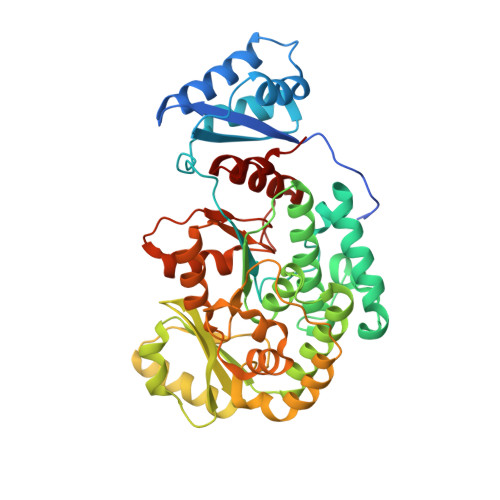
Structural Basis of a Broadly Selective Acyltransferase from the Polyketide Synthase of Splenocin.

Li, Y.Zhang, W.Zhang, H.Tian, W.Y.Wu, L.Wang, S.W.Zheng, M.M.Zhang, J.R.Sun, C.H.Deng, Z.X.Sun, Y.H.Qu, X.D.Zhou, J.H.

(2018) Angew Chem Int Ed Engl 57: 5823-5827

  • DOI: https://doi.org/10.1002/anie.201802805
  • Primary Citation Related Structures: 
    5YDA, 5YDL, 5YDM

  • PubMed Abstract: 

    Polyketides are a large family of pharmaceutically important natural products, and the structural modification of their scaffolds is significant for drug development. Herein, we report high-resolution X-ray crystal structures of the broadly selective acyltransferase (AT) from the splenocin polyketide synthase (SpnD-AT) in the apo form and in complex with benzylmalonyl and pentynylmalonyl extender unit mimics. These structures revealed the molecular basis for the stereoselectivity and substrate specificity of SpnD-AT, and enabled the engineering of the industrially important Ery-AT6 to broaden its substrate scope to include three new types of extender units.


  • Organizational Affiliation
    • Key Laboratory of Combinatorial Biosynthesis and Drug Discovery (Wuhan University), Ministry of Education, Wuhan University School of Pharmaceutical Sciences, 185 Donghu Road., Wuhan, 430071, China.

Macromolecule Content 

  • Total Structure Weight: 46.25 kDa 
  • Atom Count: 3,270 
  • Modeled Residue Count: 409 
  • Deposited Residue Count: 433 
  • Unique protein chains: 1

Macromolecules

Find similar proteins by:|  3D Structure
Entity ID: 1
MoleculeChains  Sequence LengthOrganismDetailsImage
PKS433Streptomyces sp. CNQ431Mutation(s): 0 
Gene Names: spnD
UniProt
Find proteins for A0A0E3JLZ0 (Streptomyces sp. CNQ431)
Explore A0A0E3JLZ0 
Go to UniProtKB:  A0A0E3JLZ0
Entity Groups
Sequence Clusters30% Identity50% Identity70% Identity90% Identity95% Identity100% Identity
UniProt GroupA0A0E3JLZ0
Sequence Annotations
Expand
Reference Sequence

Experimental Data & Validation

Experimental Data

  • Method: X-RAY DIFFRACTION
  • Resolution: 2.35 Å
  • R-Value Free:  0.215 (Depositor), 0.219 (DCC) 
  • R-Value Work:  0.195 (Depositor), 0.201 (DCC) 
  • R-Value Observed: 0.196 (Depositor) 
Space Group: P 43 2 2
Unit Cell:
Length ( Å )Angle ( ˚ )
a = 109.78α = 90
b = 109.78β = 90
c = 142.573γ = 90
Software Package:
Software NamePurpose
PHENIXphasing
HKL-3000data reduction
HKL-3000data scaling
PHENIXrefinement

Structure Validation

View Full Validation Report


Entry History 

& Funding Information

Deposition Data


Funding OrganizationLocationGrant Number
China--

Revision History  (Full details and data files)

  • Version 1.0: 2018-05-23
    Type: Initial release
  • Version 1.1: 2024-03-27
    Changes: Data collection, Database references