5Y78 | pdb_00005y78

Crystal structure of the triose-phosphate/phosphate translocator in complex with inorganic phosphate


Experimental Data Snapshot

  • Method: X-RAY DIFFRACTION
  • Resolution: 2.10 Å
  • R-Value Free: 
    0.226 (Depositor), 0.226 (DCC) 
  • R-Value Work: 
    0.195 (Depositor), 0.196 (DCC) 
  • R-Value Observed: 
    0.196 (Depositor) 

Starting Model: experimental
View more details

wwPDB Validation 3D Report Full Report

Validation slider image for 5Y78

Ligand Structure Quality Assessment 


This is version 1.3 of the entry. See complete history

Literature

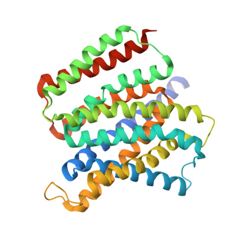
Structure of the triose-phosphate/phosphate translocator reveals the basis of substrate specificity

Lee, Y.Nishizawa, T.Takemoto, M.Kumazaki, K.Yamashita, K.Hirata, K.Minoda, A.Nagatoishi, S.Tsumoto, K.Ishitani, R.Nureki, O.

(2017) Nat Plants 3: 825-832

  • DOI: https://doi.org/10.1038/s41477-017-0022-8
  • Primary Citation Related Structures: 
    5Y78, 5Y79

  • PubMed Abstract: 

    The triose-phosphate/phosphate translocator (TPT) catalyses the strict 1:1 exchange of triose-phosphate, 3-phosphoglycerate and inorganic phosphate across the chloroplast envelope, and plays crucial roles in photosynthesis. Despite rigorous study for more than 40 years, the molecular mechanism of TPT is poorly understood because of the lack of structural information. Here we report crystal structures of TPT bound to two different substrates, 3-phosphoglycerate and inorganic phosphate, in occluded conformations. The structures reveal that TPT adopts a 10-transmembrane drug/metabolite transporter fold. Both substrates are bound within the same central pocket, where conserved lysine, arginine and tyrosine residues recognize the shared phosphate group. A structural comparison with the outward-open conformation of the bacterial drug/metabolite transporter suggests a rocker-switch motion of helix bundles, and molecular dynamics simulations support a model in which this rocker-switch motion is tightly coupled to the substrate binding, to ensure strict 1:1 exchange. These results reveal the unique mechanism of sugar phosphate/phosphate exchange by TPT.


  • Organizational Affiliation
    • Department of Biological Sciences, Graduate School of Science, University of Tokyo, 2-11-16 Yayoi, Bunkyo-ku, Tokyo, 113-0032, Japan.

Macromolecule Content 

  • Total Structure Weight: 88.32 kDa 
  • Atom Count: 5,572 
  • Modeled Residue Count: 610 
  • Deposited Residue Count: 658 
  • Unique protein chains: 1

Macromolecules

Find similar proteins by:|  3D Structure
Entity ID: 1
MoleculeChains  Sequence LengthOrganismDetailsImage
Putative hexose phosphate translocator
A, B
329Galdieria sulphurariaMutation(s): 0 
Gene Names: Gasu_48050Gs48050.1
Membrane Entity: Yes 
UniProt
Find proteins for B5AJT1 (Galdieria sulphuraria)
Explore B5AJT1 
Go to UniProtKB:  B5AJT1
Entity Groups
Sequence Clusters30% Identity50% Identity70% Identity90% Identity95% Identity100% Identity
UniProt GroupB5AJT1
Sequence Annotations
Expand
Reference Sequence

Small Molecules

Ligands 2 Unique
IDChains Name / Formula / InChI Key2D Diagram3D Interactions
OLC

Query on OLC


Download:Ideal Coordinates CCD File
AA [auth B]
BA [auth B]
CA [auth B]
D [auth A]
DA [auth B]
AA [auth B],
BA [auth B],
CA [auth B],
D [auth A],
DA [auth B],
E [auth A],
EA [auth B],
F [auth A],
FA [auth B],
G [auth A],
GA [auth B],
H [auth A],
HA [auth B],
I [auth A],
IA [auth B],
J [auth A],
JA [auth B],
K [auth A],
KA [auth B],
L [auth A],
LA [auth B],
M [auth A],
MA [auth B],
N [auth A],
NA [auth B],
O [auth A],
OA [auth B],
P [auth A],
PA [auth B],
Q [auth A],
QA [auth B],
R [auth A],
RA [auth B],
S [auth A],
SA [auth B],
T [auth A],
TA [auth B],
V [auth B],
W [auth B],
X [auth B],
Y [auth B],
Z [auth B]
(2R)-2,3-dihydroxypropyl (9Z)-octadec-9-enoate
C21 H40 O4
RZRNAYUHWVFMIP-GDCKJWNLSA-N
PO4
(Subject of Investigation/LOI)

Query on PO4


Download:Ideal Coordinates CCD File
C [auth A],
U [auth B]
PHOSPHATE ION
O4 P
NBIIXXVUZAFLBC-UHFFFAOYSA-K

Experimental Data & Validation

Experimental Data

  • Method: X-RAY DIFFRACTION
  • Resolution: 2.10 Å
  • R-Value Free:  0.226 (Depositor), 0.226 (DCC) 
  • R-Value Work:  0.195 (Depositor), 0.196 (DCC) 
  • R-Value Observed: 0.196 (Depositor) 
Space Group: P 21 21 2
Unit Cell:
Length ( Å )Angle ( ˚ )
a = 106.55α = 90
b = 164.97β = 90
c = 41.42γ = 90
Software Package:
Software NamePurpose
PHENIXrefinement
XDSdata reduction
XSCALEdata scaling
PHASERphasing

Structure Validation

View Full Validation Report


Ligand Structure Quality Assessment 


Entry History 

Deposition Data

Revision History  (Full details and data files)

  • Version 1.0: 2017-10-04
    Type: Initial release
  • Version 1.1: 2017-10-18
    Changes: Database references
  • Version 1.2: 2017-11-01
    Changes: Database references
  • Version 1.3: 2023-11-22
    Changes: Data collection, Database references, Refinement description