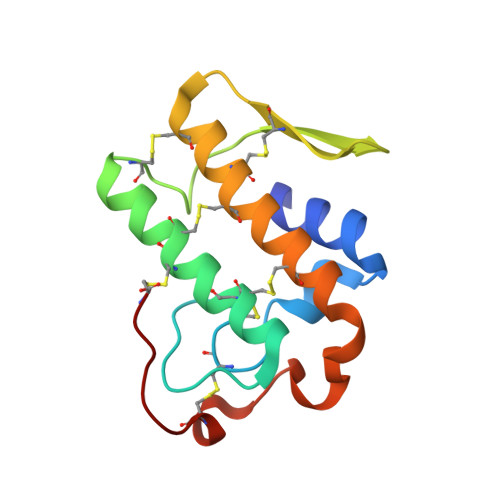
5Y5E

Crystal structure of phospholipase A2 with inhibitor


Experimental Data Snapshot

  • Method: X-RAY DIFFRACTION
  • Resolution: 1.80 Å
  • R-Value Free: 0.225 
  • R-Value Work: 0.193 
  • R-Value Observed: 0.194 

Starting Model: experimental
View more details

wwPDB Validation   3D Report Full Report


Ligand Structure Quality Assessment 


This is version 1.2 of the entry. See complete history


Literature

Structural basis for functional selectivity and ligand recognition revealed by crystal structures of human secreted phospholipase A2group IIE

Hou, S.Xu, T.Xu, J.Qu, L.Xu, Y.Chen, L.Liu, J.

(2017) Sci Rep 7: 10815-10815

  • DOI: https://doi.org/10.1038/s41598-017-11219-8
  • Primary Citation of Related Structures:  
    5WZM, 5WZO, 5WZS, 5WZT, 5WZU, 5WZV, 5WZW, 5Y5E

  • PubMed Abstract: 

    Secreted phospholipases A 2 s (sPLA 2 s) are involved in various pathological conditions such as rheumatoid arthritis and cardiovascular disease. Many inhibitors were developed and studied in clinical trials, but none have reached the market yet. This failure may be attributed to the lack of subtype selectivity for these inhibitors. Therefore, more structural information for subtype sPLA 2 is needed to guide the selective inhibitor development. In this study, the crystal structure of human sPLA 2 Group IIE (hGIIE), coupled with mutagenesis experiments, proved that the flexible second calcium binding site and residue Asn21 in hGIIE are essential to its enzymatic activity. Five inhibitor bound hGIIE complex structures revealed the key residues (Asn21 and Gly6) of hGIIE that are responsible for interacting with inhibitors, and illustrated the difference in the inhibitor binding pocket with other sPLA 2 s. This will facilitate the structure-based design of sPLA 2 's selective inhibitors.


  • Organizational Affiliation

    State Key Laboratory of Respiratory Disease, Guangzhou Institutes of Biomedicine and Health, Chinese Academy of Sciences, Guangzhou, 510530, China.


Macromolecules
Find similar proteins by:  (by identity cutoff)  |  3D Structure
Entity ID: 1
MoleculeChains Sequence LengthOrganismDetailsImage
Group IIE secretory phospholipase A2123Homo sapiensMutation(s): 1 
Gene Names: PLA2G2E
EC: 3.1.1.4
UniProt & NIH Common Fund Data Resources
Find proteins for Q9NZK7 (Homo sapiens)
Explore Q9NZK7 
Go to UniProtKB:  Q9NZK7
PHAROS:  Q9NZK7
GTEx:  ENSG00000188784 
Entity Groups  
Sequence Clusters30% Identity50% Identity70% Identity90% Identity95% Identity100% Identity
UniProt GroupQ9NZK7
Sequence Annotations
Expand
  • Reference Sequence
Small Molecules
Ligands 6 Unique
IDChains Name / Formula / InChI Key2D Diagram3D Interactions
7W3
Query on 7W3

Download Ideal Coordinates CCD File 
C [auth A]2-[2-methyl-3-oxamoyl-1-[[2-(trifluoromethyl)phenyl]methyl]indol-4-yl]oxyethanoic acid
C21 H17 F3 N2 O5
AKFUWCKNXCSWDQ-UHFFFAOYSA-N
B3P
Query on B3P

Download Ideal Coordinates CCD File 
J [auth A]2-[3-(2-HYDROXY-1,1-DIHYDROXYMETHYL-ETHYLAMINO)-PROPYLAMINO]-2-HYDROXYMETHYL-PROPANE-1,3-DIOL
C11 H26 N2 O6
HHKZCCWKTZRCCL-UHFFFAOYSA-N
GOL
Query on GOL

Download Ideal Coordinates CCD File 
B [auth A]GLYCEROL
C3 H8 O3
PEDCQBHIVMGVHV-UHFFFAOYSA-N
CA
Query on CA

Download Ideal Coordinates CCD File 
I [auth A]CALCIUM ION
Ca
BHPQYMZQTOCNFJ-UHFFFAOYSA-N
CL
Query on CL

Download Ideal Coordinates CCD File 
D [auth A],
E [auth A],
F [auth A],
G [auth A],
H [auth A]
CHLORIDE ION
Cl
VEXZGXHMUGYJMC-UHFFFAOYSA-M
NA
Query on NA

Download Ideal Coordinates CCD File 
K [auth A]SODIUM ION
Na
FKNQFGJONOIPTF-UHFFFAOYSA-N
Experimental Data & Validation

Experimental Data

  • Method: X-RAY DIFFRACTION
  • Resolution: 1.80 Å
  • R-Value Free: 0.225 
  • R-Value Work: 0.193 
  • R-Value Observed: 0.194 
  • Space Group: P 21 2 2
Unit Cell:
Length ( Å )Angle ( ˚ )
a = 48.726α = 90
b = 61.063β = 90
c = 63.356γ = 90
Software Package:
Software NamePurpose
Aimlessdata scaling
REFMACrefinement
PDB_EXTRACTdata extraction
CrysalisProdata reduction
MOLREPphasing

Structure Validation

View Full Validation Report


Ligand Structure Quality Assessment 


Entry History 

Deposition Data

Revision History  (Full details and data files)

  • Version 1.0: 2018-06-20
    Type: Initial release
  • Version 1.1: 2023-11-22
    Changes: Data collection, Database references, Refinement description
  • Version 1.2: 2024-10-23
    Changes: Structure summary