5Y2Y | pdb_00005y2y

Crystal structure of HaloTag (M175C) complexed with dansyl-PEG2-HaloTag ligand


Experimental Data Snapshot

  • Method: X-RAY DIFFRACTION
  • Resolution: 2.27 Å
  • R-Value Free: 
    0.216 (Depositor), 0.216 (DCC) 
  • R-Value Work: 
    0.158 (Depositor), 0.161 (DCC) 
  • R-Value Observed: 
    0.161 (Depositor) 

Starting Model: experimental
View more details

wwPDB Validation 3D Report Full Report

Validation slider image for 5Y2Y

Ligand Structure Quality Assessment 


This is version 1.2 of the entry. See complete history

Literature

Structure-guided synthesis of a protein-based fluorescent sensor for alkyl halides

Kang, M.G.Lee, H.Kim, B.H.Dunbayev, Y.Seo, J.K.Lee, C.Rhee, H.W.

(2017) Chem Commun (Camb) 53: 9226-9229

  • DOI: https://doi.org/10.1039/c7cc03714g
  • Primary Citation Related Structures: 
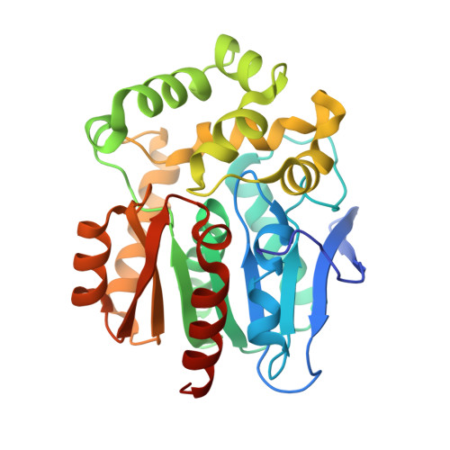
    5Y2X, 5Y2Y

  • PubMed Abstract: 

    Alkyl halides are potentially mutagenic carcinogens. However, no efficient fluorescent sensor for alkyl halide detection in human-derived samples has been developed to date. Herein, we report a new protein-based fluorescent sensor for alkyl halides. Analysis of the HaloTag holo-crystal structure with its covalently attached ligand revealed an unexpected cavity, allowing for the design of a new fluorogenic ligand. This ligand showed the highest fluorescence response (300-fold) and fastest binding kinetics (t 1/2 < 150 s) to a HaloTag mutant (M175P) protein. This protein-based sensor system was effectively used to detect alkyl halides in human serum and monitor real-time protein alkylation.


  • Organizational Affiliation
    • Department of Chemistry, Ulsan National Institute of Science and Technology (UNIST), Ulsan 44919, Korea. rhee@unist.ac.kr.

Macromolecule Content 

  • Total Structure Weight: 68.35 kDa 
  • Atom Count: 5,048 
  • Modeled Residue Count: 583 
  • Deposited Residue Count: 598 
  • Unique protein chains: 1

Macromolecules

Find similar proteins by:|  3D Structure
Entity ID: 1
MoleculeChains  Sequence LengthOrganismDetailsImage
Haloalkane dehalogenase
A, B
299Rhodococcus sp. (in: high G+C Gram-positive bacteria)Mutation(s): 22 
Gene Names: dhaA
EC: 3.8.1.5
UniProt
Find proteins for P0A3G3 (Rhodococcus sp)
Explore P0A3G3 
Go to UniProtKB:  P0A3G3
Entity Groups
Sequence Clusters30% Identity50% Identity70% Identity90% Identity95% Identity100% Identity
UniProt GroupP0A3G3
Sequence Annotations
Expand
Reference Sequence

Experimental Data & Validation

Experimental Data

  • Method: X-RAY DIFFRACTION
  • Resolution: 2.27 Å
  • R-Value Free:  0.216 (Depositor), 0.216 (DCC) 
  • R-Value Work:  0.158 (Depositor), 0.161 (DCC) 
  • R-Value Observed: 0.161 (Depositor) 
Space Group: P 21 21 21
Unit Cell:
Length ( Å )Angle ( ˚ )
a = 44.393α = 90
b = 72.355β = 90
c = 174.942γ = 90
Software Package:
Software NamePurpose
HKL-2000data processing
PHENIXphasing
Cootmodel building
PHENIXrefinement
HKL-2000data reduction
HKL-2000data scaling

Structure Validation

View Full Validation Report


Ligand Structure Quality Assessment 


Entry History 

Deposition Data

Revision History  (Full details and data files)

  • Version 1.0: 2017-09-06
    Type: Initial release
  • Version 1.1: 2023-11-22
    Changes: Advisory, Data collection, Database references, Refinement description
  • Version 1.2: 2025-09-17
    Changes: Advisory, Derived calculations, Structure summary