5XW7 | pdb_00005xw7

Crystal structure of the flexible tandem repeat domain of bacterial cellulose synthase subunit C


Experimental Data Snapshot

  • Method: X-RAY DIFFRACTION
  • Resolution: 3.27 Å
  • R-Value Free: 
    0.257 (Depositor), 0.257 (DCC) 
  • R-Value Work: 
    0.212 (Depositor), 0.216 (DCC) 
  • R-Value Observed: 
    0.215 (Depositor) 

wwPDB Validation 3D Report Full Report

Validation slider image for 5XW7

This is version 1.1 of the entry. See complete history

Literature

Crystal structure of the flexible tandem repeat domain of bacterial cellulose synthesis subunit C

Nojima, S.Fujishima, A.Kato, K.Ohuchi, K.Shimizu, N.Yonezawa, K.Tajima, K.Yao, M.

(2017) Sci Rep 7: 13018-13018

  • DOI: https://doi.org/10.1038/s41598-017-12530-0
  • Primary Citation Related Structures: 
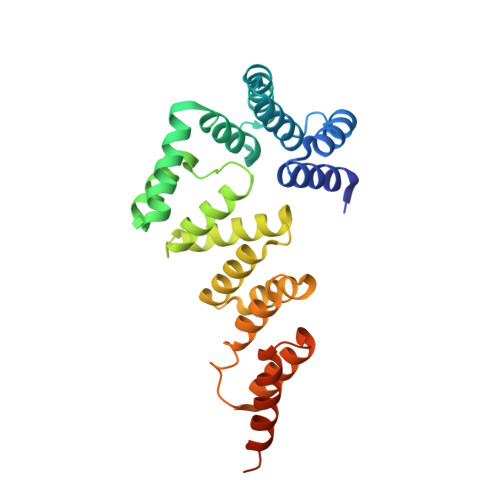
    5XW7

  • PubMed Abstract: 

    Bacterial cellulose (BC) is synthesized and exported through the cell membrane via a large protein complex (terminal complex) that consists of three or four subunits. BcsC is a little-studied subunit considered to export BC to the extracellular matrix. It is predicted to have two domains: a tetratrico peptide repeat (TPR) domain and a β-barrelled outer membrane domain. Here we report the crystal structure of the N-terminal part of BcsC-TPR domain (Asp24-Arg272) derived from Enterobacter CJF-002. Unlike most TPR-containing proteins which have continuous TPR motifs, this structure has an extra α-helix between two clusters of TPR motifs. Five independent molecules in the crystal had three different conformations that varied at the hinge of the inserted α-helix. Such structural feature indicates that the inserted α-helix confers flexibility to the chain and changes the direction of the TPR super-helix, which was also suggested by structural analysis of BcsC-TPR (Asp24-Leu664) in solution by size exclusion chromatography-small-angle X-ray scattering. The flexibility at the α-helical hinge may play important role for exporting glucan chains.


  • Organizational Affiliation
    • Graduate school of Life Science, Hokkaido University, Sapporo, Hokkaido, 060-0810, Japan.

Macromolecule Content 

  • Total Structure Weight: 142.77 kDa 
  • Atom Count: 8,740 
  • Modeled Residue Count: 1,146 
  • Deposited Residue Count: 1,300 
  • Unique protein chains: 1

Macromolecules

Find similar proteins by:|  3D Structure
Entity ID: 1
MoleculeChains  Sequence LengthOrganismDetailsImage
Cellulose synthase subunit C
A, B, C, D, E
260Enterobacter sp. CJF-002Mutation(s): 0 
Gene Names: bcsC
UniProt
Find proteins for K0J1W8 (Enterobacter sp. CJF-002)
Explore K0J1W8 
Go to UniProtKB:  K0J1W8
Entity Groups
Sequence Clusters30% Identity50% Identity70% Identity90% Identity95% Identity100% Identity
UniProt GroupK0J1W8
Sequence Annotations
Expand
Reference Sequence

Experimental Data & Validation

Experimental Data

  • Method: X-RAY DIFFRACTION
  • Resolution: 3.27 Å
  • R-Value Free:  0.257 (Depositor), 0.257 (DCC) 
  • R-Value Work:  0.212 (Depositor), 0.216 (DCC) 
  • R-Value Observed: 0.215 (Depositor) 
Space Group: P 1 21 1
Unit Cell:
Length ( Å )Angle ( ˚ )
a = 115.81α = 90
b = 55.62β = 95.56
c = 165.02γ = 90
Software Package:
Software NamePurpose
PHENIXrefinement
XDSdata scaling
PHENIXphasing
XDSdata reduction

Structure Validation

View Full Validation Report


Entry History 

Deposition Data

Revision History  (Full details and data files)

  • Version 1.0: 2017-11-01
    Type: Initial release
  • Version 1.1: 2024-03-27
    Changes: Data collection, Database references