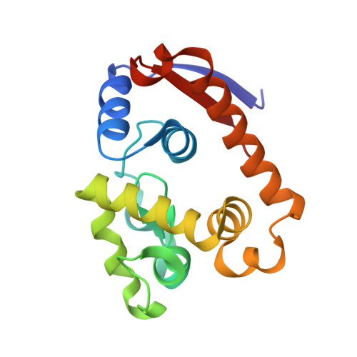
5XU5

Complex structure of RmMan134A-M4


Experimental Data Snapshot

  • Method: X-RAY DIFFRACTION
  • Resolution: 2.02 Å
  • R-Value Free: 0.215 
  • R-Value Work: 0.156 

wwPDB Validation   3D Report Full Report


This is version 2.2 of the entry. See complete history


Literature

Structural and biochemical insights into the substrate-binding mechanism of a novel glycoside hydrolase family 134 beta-mannanase.

You, X.Qin, Z.Li, Y.X.Yan, Q.J.Li, B.Jiang, Z.Q.

(2018) Biochim Biophys Acta Gen Subj 1862: 1376-1388

  • DOI: https://doi.org/10.1016/j.bbagen.2018.03.016
  • Primary Citation of Related Structures:  
    5XTJ, 5XTT, 5XU5, 5XUG, 5XUL

  • PubMed Abstract: 

    Mannan is one of the major constituent groups of hemicellulose, which is a renewable resource from higher plants. β-Mannanases are enzymes capable of degrading lignocellulosic biomass. Here, an endo-β-mannanase from Rhizopus microsporus (RmMan134A) was cloned and expressed. The recombinant RmMan134A showed maximal activity at pH 5.0 and 50 °C, and exhibited high specific activity towards locust bean gum (2337 U/mg). To gain insight into the substrate-binding mechanism of RmMan134A, four complex structures (RmMan134A-M3, RmMan134A-M4, RmMan134A-M5 and RmMan134A-M6) were further solved. These structures showed that there were at least seven subsites (-3 to +4) in the catalytic groove of RmMan134A. Mannose in the -1 subsite hydrogen bonded with His113 and Tyr131, revealing a unique conformation. Lys48 and Val159 formed steric hindrance, which impedes to bond with galactose branches. In addition, the various binding modes of RmMan134A-M5 indicated that subsites -2 to +2 are indispensable during the hydrolytic process. The structure of RmMan134A-M4 showed that mannotetrose only binds at subsites +1 to +4, and RmMan134A could therefore not hydrolyze mannan oligosaccharides with degree of polymerization ≤4. Through rational design, the specific activity and optimal conditions of RmMan134A were significantly improved. The purpose of this paper is to investigate the structure and function of fungal GH family 134 β-1,4-mannanases, and substrate-binding mechanism of GH family 134 members.


  • Organizational Affiliation

    Beijing Advanced Innovation Center for Food Nutrition and Human Health, College of Engineering, China Agricultural University, Beijing 100083, China.


Macromolecules
Find similar proteins by:  (by identity cutoff)  |  3D Structure
Entity ID: 1
MoleculeChains Sequence LengthOrganismDetailsImage
endo-1,4-beta-mannanaseA [auth B],
B [auth A]
162Rhizopus microsporusMutation(s): 0 
Gene Names: BCV71DRAFT_26579RMCBS344292_04886
EC: 3.2.1.78
UniProt
Find proteins for A0A2U8ZTY7 (Rhizopus microsporus var. rhizopodiformis)
Explore A0A2U8ZTY7 
Go to UniProtKB:  A0A2U8ZTY7
Entity Groups  
Sequence Clusters30% Identity50% Identity70% Identity90% Identity95% Identity100% Identity
UniProt GroupA0A2U8ZTY7
Sequence Annotations
Expand
  • Reference Sequence
Oligosaccharides

Help

Entity ID: 2
MoleculeChains Length2D Diagram Glycosylation3D Interactions
beta-D-mannopyranose-(1-4)-beta-D-mannopyranose-(1-4)-beta-D-mannopyranose-(1-4)-beta-D-mannopyranose
C, D
4N/A
Glycosylation Resources
GlyTouCan:  G51338PC
GlyCosmos:  G51338PC
GlyGen:  G51338PC
Experimental Data & Validation

Experimental Data

  • Method: X-RAY DIFFRACTION
  • Resolution: 2.02 Å
  • R-Value Free: 0.215 
  • R-Value Work: 0.156 
  • Space Group: P 21 21 21
Unit Cell:
Length ( Å )Angle ( ˚ )
a = 44.696α = 90
b = 59.002β = 90
c = 104.983γ = 90
Software Package:
Software NamePurpose
PHENIXrefinement
HKL-3000data reduction
HKL-3000data scaling
HKL-3000phasing

Structure Validation

View Full Validation Report


Entry History 

Deposition Data

Revision History  (Full details and data files)

  • Version 1.0: 2018-06-27
    Type: Initial release
  • Version 1.1: 2019-07-10
    Changes: Data collection, Database references
  • Version 2.0: 2020-07-29
    Type: Remediation
    Reason: Carbohydrate remediation
    Changes: Atomic model, Data collection, Derived calculations, Structure summary
  • Version 2.1: 2020-09-09
    Changes: Structure summary
  • Version 2.2: 2024-03-27
    Changes: Data collection, Database references