5XU0 | pdb_00005xu0

Structure of the membrane fusion protein Spr0693 from Streptococcus pneumoniae R6


Experimental Data Snapshot

  • Method: X-RAY DIFFRACTION
  • Resolution: 2.95 Å
  • R-Value Free: 
    0.305 (Depositor), 0.305 (DCC) 
  • R-Value Work: 
    0.248 (Depositor) 
  • R-Value Observed: 
    0.250 (Depositor) 

Starting Model: experimental
View more details

wwPDB Validation 3D Report Full Report

Validation slider image for 5XU0

This is version 1.3 of the entry. See complete history

Literature

Structure of a MacAB-like efflux pump from Streptococcus pneumoniae.

Yang, H.B.Hou, W.T.Cheng, M.T.Jiang, Y.L.Chen, Y.Zhou, C.Z.

(2018) Nat Commun 9: 196-196

  • DOI: https://doi.org/10.1038/s41467-017-02741-4
  • Primary Citation Related Structures: 
    5XU0, 5XU1

  • PubMed Abstract: 

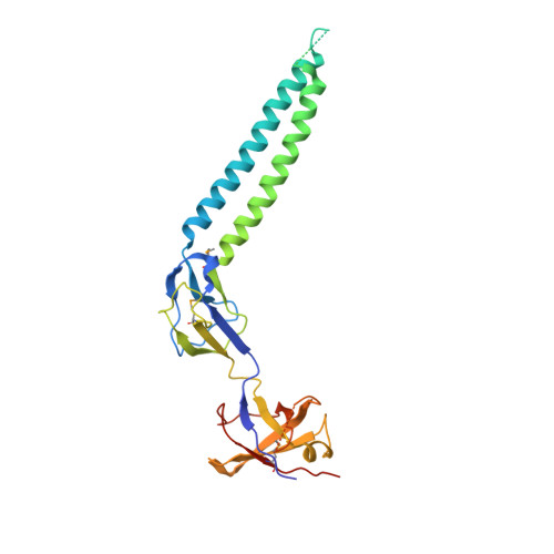
    The spr0693-spr0694-spr0695 operon of Streptococcus pneumoniae encodes a putative ATP-binding cassette (ABC)-type efflux pump involved in the resistance of antibiotics and antimicrobial peptides. Here we report the crystal structures of Spr0694-0695 at 3.3 Å and Spr0693 at 3.0 Å resolution, revealing a MacAB-like efflux pump. The dimeric Spr0694-0695 adopts a non-canonical fold of ABC transporter, the transmembrane domain of which consists of eight tightly packed transmembrane helices with an insertion of extracellular domain between the first and second helices, whereas Spr0693 forms a nanotube channel docked onto the ABC transporter. Structural analyses combined with ATPase activity and antimicrobial susceptibility assays, enable us to propose a putative substrate-entrance tunnel with a lateral access controlled by a guard helix. Altogether, our findings provide structural insights and putative transport mechanism of a MacAB-like efflux pump in Gram-positive bacteria.


  • Organizational Affiliation
    • Hefei National Laboratory for Physical Sciences at the Microscale and School of Life Sciences, University of Science and Technology of China, Hefei, Anhui, 230027, China.

Macromolecule Content 

  • Total Structure Weight: 87.07 kDa 
  • Atom Count: 5,102 
  • Modeled Residue Count: 681 
  • Deposited Residue Count: 819 
  • Unique protein chains: 1

Macromolecules

Find similar proteins by:|  3D Structure
Entity ID: 1
MoleculeChains  Sequence LengthOrganismDetailsImage
Membrane-fusion protein
A, B, C
273Streptococcus pneumoniaeMutation(s): 2 
Gene Names: ERS515107_03100

Small Molecules

Modified Residues  1 Unique
IDChains TypeFormula2D DiagramParent
MSE
Query on MSE
A, B, C
L-PEPTIDE LINKINGC5 H11 N O2 SeMET

Experimental Data & Validation

Experimental Data

  • Method: X-RAY DIFFRACTION
  • Resolution: 2.95 Å
  • R-Value Free:  0.305 (Depositor), 0.305 (DCC) 
  • R-Value Work:  0.248 (Depositor) 
  • R-Value Observed: 0.250 (Depositor) 
Space Group: P 41 21 2
Unit Cell:
Length ( Å )Angle ( ˚ )
a = 159.21α = 90
b = 159.21β = 90
c = 99.4γ = 90
Software Package:
Software NamePurpose
PHENIXrefinement
XDSdata reduction
XSCALEdata scaling
MOLREPphasing

Structure Validation

View Full Validation Report


Entry History 

& Funding Information

Deposition Data


Funding OrganizationLocationGrant Number
Ministry of Science and TechnologyChina2015CB910103
National Natural Science Foundation of ChinaChina31400628

Revision History  (Full details and data files)

  • Version 1.0: 2018-01-24
    Type: Initial release
  • Version 1.1: 2018-01-31
    Changes: Database references
  • Version 1.2: 2023-11-22
    Changes: Data collection, Database references, Refinement description
  • Version 1.3: 2024-10-30
    Changes: Structure summary