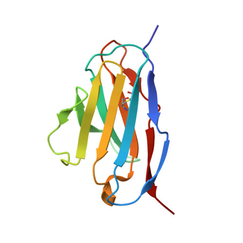
5XP1

Structure of monomeric mutant of REI immunoglobulin light chain variable domain crystallized at pH 6


Experimental Data Snapshot

  • Method: X-RAY DIFFRACTION
  • Resolution: 2.88 Å
  • R-Value Free: 0.297 
  • R-Value Work: 0.257 
  • R-Value Observed: 0.259 

Starting Model: experimental
View more details

wwPDB Validation   3D Report Full Report


This is version 1.3 of the entry. See complete history

Re-refinement Note

This entry reflects an alternative modeling of the original data in: 5B3C


Literature

Heat-induced native dimerization prevents amyloid formation by variable domain from immunoglobulin light-chain REI

Nawata, M.Tsutsumi, H.Kobayashi, Y.Unzai, S.Mine, S.Nakamura, T.Uegaki, K.Kamikubo, H.Kataoka, M.Hamada, D.

(2017) FEBS J 284: 3114-3127

  • DOI: https://doi.org/10.1111/febs.14181
  • Primary Citation of Related Structures:  
    5XP1, 5XQY

  • PubMed Abstract: 

    Amyloid light-chain (AL) amyloidosis is a protein-misfolding disease characterized by accumulation of immunoglobulin light chains (LCs) into amyloid fibrils. Dimerization of a full length or variable domain (V L ) of LC serves to stabilize the native state and prevent the formation of amyloid fibrils. We here analyzed the thermodynamic properties of dimerization and unfolding reactions by nonamyloidogenic V L from REI LC or its monomeric Y96K mutant using sedimentation velocity and circular dichroism. The data indicate that the equilibrium shifts to native dimerization for wild-type REI V L by elevating temperature due to the negative enthalpy change for dimer dissociation (-81.2 kJ·mol -1 ). The Y96K mutation did not affect the stability of the monomeric native state but increased amyloidogenicity. These results suggest that the heat-induced native homodimerization is the major factor preventing amyloid formation by wild-type REI V L . Heat-induced native oligomerization may be an efficient strategy to avoid the formation of misfolded aggregates particularly for thermostable proteins that are used at elevated temperatures under conditions where other proteins tend to misfold. Structural data are available in the Protein Data Bank under the accession numbers 5XP1 and 5XQY.


  • Organizational Affiliation

    Graduate School of Medicine, Kobe University, Japan.


Macromolecules
Find similar proteins by:  (by identity cutoff)  |  3D Structure
Entity ID: 1
MoleculeChains Sequence LengthOrganismDetailsImage
Immunoglobulin kappa variable 1D-33
A, B, C, D, E
A, B, C, D, E, F, G, H
109Homo sapiensMutation(s): 0 
Gene Names: IGKV1D-33
UniProt & NIH Common Fund Data Resources
Find proteins for P01593 (Homo sapiens)
Explore P01593 
Go to UniProtKB:  P01593
GTEx:  ENSG00000239975 
Entity Groups  
Sequence Clusters30% Identity50% Identity70% Identity90% Identity95% Identity100% Identity
UniProt GroupP01593
Sequence Annotations
Expand
  • Reference Sequence
Experimental Data & Validation

Experimental Data

  • Method: X-RAY DIFFRACTION
  • Resolution: 2.88 Å
  • R-Value Free: 0.297 
  • R-Value Work: 0.257 
  • R-Value Observed: 0.259 
  • Space Group: P 4
Unit Cell:
Length ( Å )Angle ( ˚ )
a = 130.76α = 90
b = 130.76β = 90
c = 92.14γ = 90
Software Package:
Software NamePurpose
PHENIXrefinement
iMOSFLMdata reduction
SCALAdata scaling
MOLREPphasing

Structure Validation

View Full Validation Report


Entry History & Funding Information

Deposition Data


Funding OrganizationLocationGrant Number
JSPSJapan26440076
JSPSJapan23107721

Revision History  (Full details and data files)

  • Version 1.0: 2017-08-02
    Type: Initial release
  • Version 1.1: 2017-10-25
    Changes: Database references
  • Version 1.2: 2023-11-22
    Changes: Data collection, Database references, Refinement description
  • Version 1.3: 2024-11-20
    Changes: Structure summary