5XOY

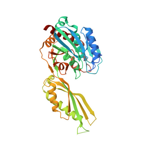
Crystal structure of LysK from Thermus thermophilus in complex with Lysine


Experimental Data Snapshot

  • Method: X-RAY DIFFRACTION
  • Resolution: 2.39 Å
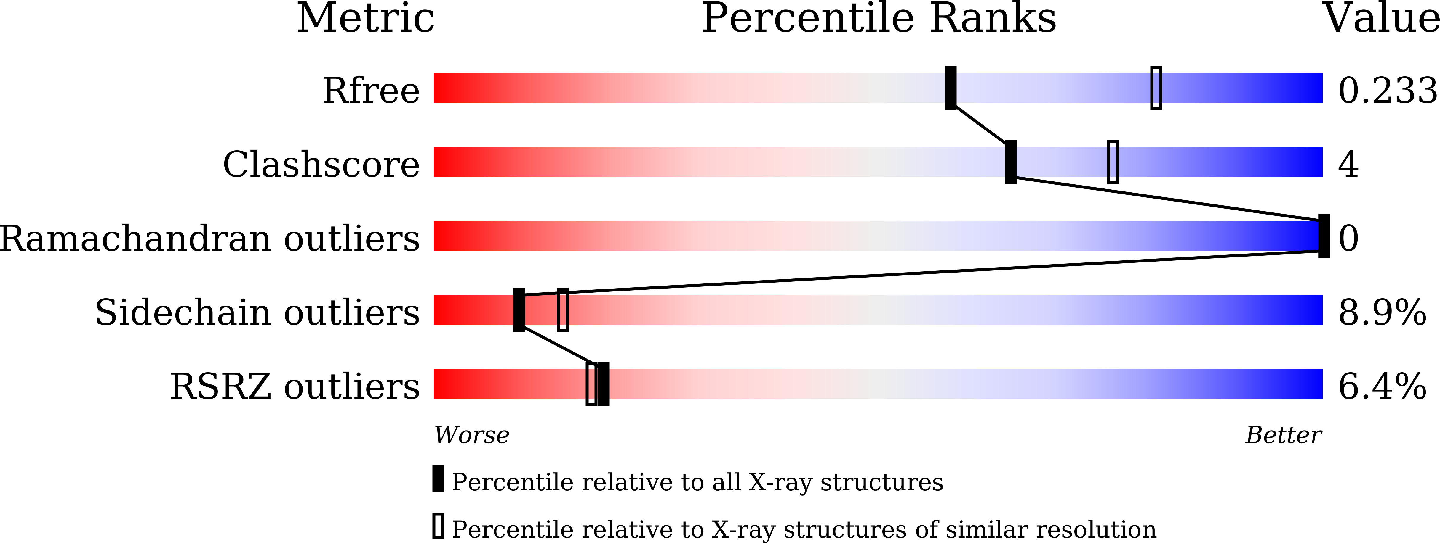
  • R-Value Free: 0.229 
  • R-Value Work: 0.188 
  • R-Value Observed: 0.190 

Starting Model: experimental
View more details

wwPDB Validation   3D Report Full Report


This is version 1.1 of the entry. See complete history


Literature

Crystal structure of LysK, an enzyme catalyzing the last step of lysine biosynthesis in Thermus thermophilus, in complex with lysine: Insight into the mechanism for recognition of the amino-group carrier protein, LysW

Fujita, S.Cho, S.-H.Yoshida, A.Hasebe, F.Tomita, T.Kuzuyama, T.Nishiyama, M.

(2017) Biochem Biophys Res Commun 491: 409-415

  • DOI: https://doi.org/10.1016/j.bbrc.2017.07.088
  • Primary Citation of Related Structures:  
    5XOY

  • PubMed Abstract: 

    LysK is an M20 peptidase family enzyme that hydrolyzes the isopeptide bond between the carrier protein LysW and lysine in order to release lysine, which is the last step of lysine biosynthesis in Thermus thermophilus. In the present study, we determined the crystal structure of LysK in complex with lysine at a resolution of 2.4 Å. The α-amino group of the bound lysine was oriented toward the catalytic center, which was composed of the residues coordinating divalent metal ions for the hydrolysis of the isopeptide bond. An 11 Å-long path was observed from the active site binding lysine to the protein surface, which may be responsible for recognizing the C-terminal extension domain of LysW with the conserved EDWGE sequence. A positively-charged surface region was detected around the exit of the path, similar to other lysine biosynthetic enzymes using LysW as the carrier protein. Mutational studies of the surface residues provided a plausible model for the electrostatic interaction with LysW.


  • Organizational Affiliation

    Biotechnology Research Center, The University of Tokyo, Japan.


Macromolecules
Find similar proteins by:  (by identity cutoff)  |  3D Structure
Entity ID: 1
MoleculeChains Sequence LengthOrganismDetailsImage
[LysW]-lysine hydrolase
A, B
373Thermus thermophilus HB27Mutation(s): 0 
Gene Names: lysKTT_C1396
EC: 3.5.1 (PDB Primary Data), 3.5.1.130 (UniProt)
UniProt
Find proteins for Q8VUS5 (Thermus thermophilus (strain ATCC BAA-163 / DSM 7039 / HB27))
Explore Q8VUS5 
Go to UniProtKB:  Q8VUS5
Entity Groups  
Sequence Clusters30% Identity50% Identity70% Identity90% Identity95% Identity100% Identity
UniProt GroupQ8VUS5
Sequence Annotations
Expand
  • Reference Sequence
Experimental Data & Validation

Experimental Data

  • Method: X-RAY DIFFRACTION
  • Resolution: 2.39 Å
  • R-Value Free: 0.229 
  • R-Value Work: 0.188 
  • R-Value Observed: 0.190 
  • Space Group: P 21 21 2
Unit Cell:
Length ( Å )Angle ( ˚ )
a = 132.778α = 90
b = 69.927β = 90
c = 118.094γ = 90
Software Package:
Software NamePurpose
REFMACrefinement
HKL-2000data reduction
HKL-2000data scaling
PHASERphasing

Structure Validation

View Full Validation Report


Entry History & Funding Information

Deposition Data


Funding OrganizationLocationGrant Number
JSPSJapan24228001

Revision History  (Full details and data files)

  • Version 1.0: 2017-09-13
    Type: Initial release
  • Version 1.1: 2023-11-22
    Changes: Data collection, Database references, Refinement description