5XO7 | pdb_00005xo7

Crystal structure of a novel ZEN lactonase mutant with ligand a


Experimental Data Snapshot

  • Method: X-RAY DIFFRACTION
  • Resolution: 1.88 Å
  • R-Value Free: 
    0.195 (Depositor), 0.191 (DCC) 
  • R-Value Work: 
    0.156 (Depositor) 
  • R-Value Observed: 
    0.156 (Depositor) 

Starting Model: experimental
View more details

wwPDB Validation 3D Report Full Report

Validation slider image for 5XO7

Ligand Structure Quality Assessment 


This is version 1.1 of the entry. See complete history

Literature

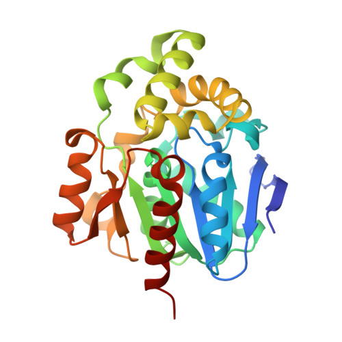
Crystal Structure of a Mycoestrogen-Detoxifying Lactonase from Rhinocladiella mackenziei: Molecular Insight into ZHD Substrate Selectivity

Zheng, Y.Y.Liu, W.T.Chen, C.C.Hu, X.Y.Liu, W.D.Ko, T.P.Tang, X.K.Wei, H.L.Huang, J.W.Guo, R.T.

(2018) ACS Catal 8: 4294-4298

Macromolecule Content 

  • Total Structure Weight: 238.16 kDa 
  • Atom Count: 18,834 
  • Modeled Residue Count: 2,104 
  • Deposited Residue Count: 2,128 
  • Unique protein chains: 1

Macromolecules

Find similar proteins by:|  3D Structure
Entity ID: 1
MoleculeChains  Sequence LengthOrganismDetailsImage
lactonase for protein
A, B, C, D, E
A, B, C, D, E, F, G, H
266Rhinocladiella mackenziei CBS 650.93Mutation(s): 1 
Gene Names: Z518_04590
UniProt
Find proteins for A0A0D2ILK1 (Rhinocladiella mackenziei CBS 650.93)
Explore A0A0D2ILK1 
Go to UniProtKB:  A0A0D2ILK1
Entity Groups
Sequence Clusters30% Identity50% Identity70% Identity90% Identity95% Identity100% Identity
UniProt GroupA0A0D2ILK1
Sequence Annotations
Expand
Reference Sequence

Small Molecules

Ligands 1 Unique
IDChains Name / Formula / InChI Key2D Diagram3D Interactions
36J

Query on 36J


Download:Ideal Coordinates CCD File
I [auth A]
J [auth B]
K [auth C]
L [auth D]
M [auth E]
I [auth A],
J [auth B],
K [auth C],
L [auth D],
M [auth E],
N [auth F],
O [auth G],
P [auth H]
(3S,7R,11E)-7,14,16-trihydroxy-3-methyl-3,4,5,6,7,8,9,10-octahydro-1H-2-benzoxacyclotetradecin-1-one
C18 H24 O5
FPQFYIAXQDXNOR-QDKLYSGJSA-N

Experimental Data & Validation

Experimental Data

  • Method: X-RAY DIFFRACTION
  • Resolution: 1.88 Å
  • R-Value Free:  0.195 (Depositor), 0.191 (DCC) 
  • R-Value Work:  0.156 (Depositor) 
  • R-Value Observed: 0.156 (Depositor) 
Space Group: P 1
Unit Cell:
Length ( Å )Angle ( ˚ )
a = 75.462α = 90.2
b = 95.158β = 92.13
c = 101.444γ = 91.6
Software Package:
Software NamePurpose
REFMACrefinement
HKL-2000data scaling
PDB_EXTRACTdata extraction
HKL-2000data reduction
PHASERphasing

Structure Validation

View Full Validation Report


Ligand Structure Quality Assessment 


Entry History 

Deposition Data

Revision History  (Full details and data files)

  • Version 1.0: 2018-05-02
    Type: Initial release
  • Version 1.1: 2023-11-22
    Changes: Data collection, Database references, Refinement description