5XM3

Crystal Structure of Methanol dehydrogenase from Methylophaga aminisulfidivorans


Experimental Data Snapshot

  • Method: X-RAY DIFFRACTION
  • Resolution: 1.70 Å
  • R-Value Free: 0.181 
  • R-Value Work: 0.151 
  • R-Value Observed: 0.153 

Starting Model: experimental
View more details

wwPDB Validation   3D Report Full Report


Ligand Structure Quality Assessment 


This is version 1.2 of the entry. See complete history


Literature

The crystal structure of methanol dehydrogenase, a quinoprotein from the marine methylotrophic bacterium Methylophaga aminisulfidivorans MPT

Cao, T.P.Choi, J.M.Kim, S.W.Lee, S.H.

(2018) J Microbiol 56: 246-254

  • DOI: https://doi.org/10.1007/s12275-018-7483-y
  • Primary Citation of Related Structures:  
    5XM3

  • PubMed Abstract: 

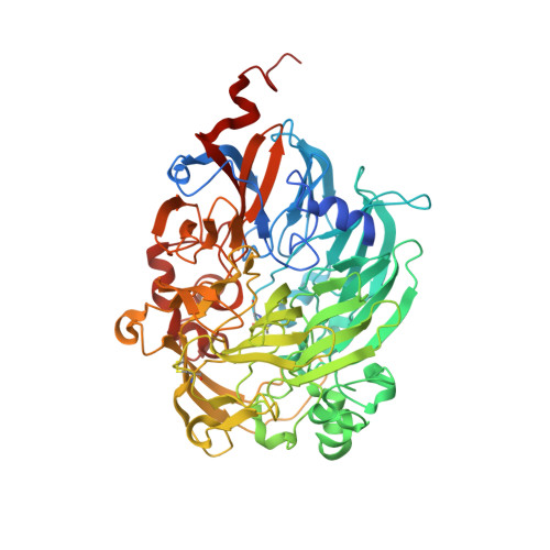
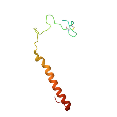
    The first crystal structure of a pyrroloquinoline quinone (PQQ)-dependent methanol dehydrogenase (MDH) from a marine methylotrophic bacterium, Methylophaga aminisulfidivorans MP T (MDH Mas ), was determined at 1.7 Å resolution. The active form of MDH Mas (or MDHI Mas ) is a heterotetrameric α 2 β 2 , where each β-subunit assembles on one side of each of the α-subunits, in a symmetrical fashion, so that two β-subunits surround the two PQQ-binding pockets on the α-subunits. The active site consists of a PQQ molecule surrounded by a β-propeller fold for each α-subunit. Interestingly, the PQQ molecules are coordinated by a Mg 2+ ion, instead of the Ca 2+ ion that is commonly found in the terrestrial MDHI, indicating the efficiency of osmotic balance regulation in the high salt environment. The overall interaction of the β-subunits with the α-subunits appears tighter than that of terrestrial homologues, suggesting the efficient maintenance of MDHI Mas integrity in the sea water environment to provide a firm basis for complex formation with MxaJ Mas or Cyt c L . With the help of the features mentioned above, our research may enable the elucidation of the full molecular mechanism of methanol oxidation by taking advantage of marine bacterium-originated proteins in the methanol oxidizing system (mox), including MxaJ, as the attainment of these proteins from terrestrial bacteria for structural studies has not been successful.


  • Organizational Affiliation

    Department of Cellular and Molecular Medicine, Chosun University School of Medicine, Gwangju, 61452, Republic of Korea.


Macromolecules
Find similar proteins by:  (by identity cutoff)  |  3D Structure
Entity ID: 1
MoleculeChains Sequence LengthOrganismDetailsImage
Glucose dehydrogenase
A, C
627Methylophaga aminisulfidivorans MPMutation(s): 0 
Gene Names: MAMP_01209
UniProt
Find proteins for A3FJ48 (Methylophaga aminisulfidivorans MP)
Explore A3FJ48 
Go to UniProtKB:  A3FJ48
Entity Groups  
Sequence Clusters30% Identity50% Identity70% Identity90% Identity95% Identity100% Identity
UniProt GroupA3FJ48
Sequence Annotations
Expand
  • Reference Sequence
Find similar proteins by:  (by identity cutoff)  |  3D Structure
Entity ID: 2
MoleculeChains Sequence LengthOrganismDetailsImage
Methanol dehydrogenase [cytochrome c] subunit 2
B, D
90Methylophaga aminisulfidivorans MPMutation(s): 0 
Gene Names: MAMP_01202
EC: 1.1.2.7
UniProt
Find proteins for A3FJ51 (Methylophaga aminisulfidivorans MP)
Explore A3FJ51 
Go to UniProtKB:  A3FJ51
Entity Groups  
Sequence Clusters30% Identity50% Identity70% Identity90% Identity95% Identity100% Identity
UniProt GroupA3FJ51
Sequence Annotations
Expand
  • Reference Sequence
Experimental Data & Validation

Experimental Data

  • Method: X-RAY DIFFRACTION
  • Resolution: 1.70 Å
  • R-Value Free: 0.181 
  • R-Value Work: 0.151 
  • R-Value Observed: 0.153 
  • Space Group: P 1 21 1
Unit Cell:
Length ( Å )Angle ( ˚ )
a = 63.926α = 90
b = 109.479β = 100.48
c = 95.554γ = 90
Software Package:
Software NamePurpose
PHENIXrefinement
HKL-2000data processing
MOLREPphasing
Cootmodel building

Structure Validation

View Full Validation Report


Ligand Structure Quality Assessment 


Entry History & Funding Information

Deposition Data


Funding OrganizationLocationGrant Number
Chosun UniversityKorea, Republic Of2015

Revision History  (Full details and data files)

  • Version 1.0: 2018-03-21
    Type: Initial release
  • Version 1.1: 2018-04-11
    Changes: Data collection, Database references, Structure summary
  • Version 1.2: 2023-11-22
    Changes: Data collection, Database references, Derived calculations, Refinement description