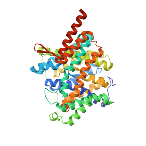
5XLS

Crystal structure of UraA in occluded conformation


Experimental Data Snapshot

  • Method: X-RAY DIFFRACTION
  • Resolution: 2.50 Å
  • R-Value Free: 0.247 
  • R-Value Work: 0.226 
  • R-Value Observed: 0.227 

Starting Model: experimental
View more details

wwPDB Validation   3D Report Full Report


This is version 1.2 of the entry. See complete history


Literature

Dimeric structure of the uracil:proton symporter UraA provides mechanistic insights into the SLC4/23/26 transporters

Yu, X.Yang, G.Yan, C.Baylon, J.L.Jiang, J.Fan, H.Lu, G.Hasegawa, K.Okumura, H.Wang, T.Tajkhorshid, E.Li, S.Yan, N.

(2017) Cell Res 27: 1020-1033

  • DOI: https://doi.org/10.1038/cr.2017.83
  • Primary Citation of Related Structures:  
    5XLS

  • PubMed Abstract: 

    The Escherichia coli uracil:proton symporter UraA is a prototypical member of the nucleobase/ascorbate transporter (NAT) or nucleobase/cation symporter 2 (NCS2) family, which corresponds to the human solute carrier family SLC23. UraA consists of 14 transmembrane segments (TMs) that are organized into two distinct domains, the core domain and the gate domain, a structural fold that is also shared by the SLC4 and SLC26 transporters. Here we present the crystal structure of UraA bound to uracil in an occluded state at 2.5 Å resolution. Structural comparison with the previously reported inward-open UraA reveals pronounced relative motions between the core domain and the gate domain as well as intra-domain rearrangement of the gate domain. The occluded UraA forms a dimer in the structure wherein the gate domains are sandwiched by two core domains. In vitro and in vivo biochemical characterizations show that UraA is at equilibrium between dimer and monomer in all tested detergent micelles, while dimer formation is necessary for the transport activity. Structural comparison between the dimeric UraA and the recently reported inward-facing dimeric UapA provides important insight into the transport mechanism of SLC23 transporters.


  • Organizational Affiliation

    State Key Laboratory of Membrane Biology, School of Life Sciences and School of Medicine, Tsinghua University, Beijing 100084, China.


Macromolecules
Find similar proteins by:  (by identity cutoff)  |  3D Structure
Entity ID: 1
MoleculeChains Sequence LengthOrganismDetailsImage
Uracil permease435Escherichia coli O157:H7Mutation(s): 0 
Gene Names: uraAZ3760ECs3359
Membrane Entity: Yes 
UniProt
Find proteins for P0AGM8 (Escherichia coli O157:H7)
Explore P0AGM8 
Go to UniProtKB:  P0AGM8
Entity Groups  
Sequence Clusters30% Identity50% Identity70% Identity90% Identity95% Identity100% Identity
UniProt GroupP0AGM8
Sequence Annotations
Expand
  • Reference Sequence
Experimental Data & Validation

Experimental Data

  • Method: X-RAY DIFFRACTION
  • Resolution: 2.50 Å
  • R-Value Free: 0.247 
  • R-Value Work: 0.226 
  • R-Value Observed: 0.227 
  • Space Group: C 2 2 21
Unit Cell:
Length ( Å )Angle ( ˚ )
a = 74.721α = 90
b = 134.726β = 90
c = 143.452γ = 90
Software Package:
Software NamePurpose
PHENIXrefinement
HKL-2000data reduction
HKL-2000data scaling
PHASERphasing

Structure Validation

View Full Validation Report


Entry History & Funding Information

Deposition Data


Funding OrganizationLocationGrant Number
Ministry of Science and Technology of ChinaChina2015CB910101, 2016YFA0500402, 2014ZX09507003-006
National Natural Science Foundation of ChinaChina--

Revision History  (Full details and data files)

  • Version 1.0: 2017-07-05
    Type: Initial release
  • Version 1.1: 2017-08-16
    Changes: Database references
  • Version 1.2: 2023-11-22
    Changes: Data collection, Database references, Refinement description