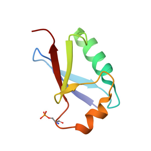
Ubiquitin S65 phosphorylation engenders a pH-sensitive conformational switch
Dong, X., Gong, Z., Lu, Y.B., Liu, K., Qin, L.Y., Ran, M.L., Zhang, C.L., Liu, Z., Zhang, W.P., Tang, C.(2017) Proc Natl Acad Sci U S A 114: 6770-6775
- PubMed: 28611216
- DOI: https://doi.org/10.1073/pnas.1705718114
- Primary Citation of Related Structures:
5XK4, 5XK5 - PubMed Abstract:
Ubiquitin (Ub) is an important signaling protein. Recent studies have shown that Ub can be enzymatically phosphorylated at S65, and that the resulting pUb exhibits two conformational states-a relaxed state and a retracted state. However, crystallization efforts have yielded only the structure for the relaxed state, which was found similar to that of unmodified Ub. Here we present the solution structures of pUb in both states obtained through refinement against state-specific NMR restraints. We show that the retracted state differs from the relaxed state by the retraction of the last β-strand and by the extension of the second α-helix. Further, we show that at 7.2, the pK a value for the phosphoryl group in the relaxed state is higher by 1.4 units than that in the retracted state. Consequently, pUb exists in equilibrium between protonated and deprotonated forms and between retracted and relaxed states, with protonated/relaxed species enriched at slightly acidic pH and deprotonated/retracted species enriched at slightly basic pH. The heterogeneity of pUb explains the inability of phosphomimetic mutants to fully mimic pUb. The pH-sensitive conformational switch is likely preserved for polyubiquitin, as single-molecule FRET data indicate that pH change leads to quaternary rearrangement of a phosphorylated K63-linked diubiquitin. Because cellular pH varies among compartments and changes upon pathophysiological insults, our finding suggests that pH and Ub phosphorylation confer additional target specificities and enable an additional layer of modulation for Ub signals.
- Key Laboratory of Magnetic Resonance in Biological Systems of the Chinese Academy of Sciences, State Key Laboratory of Magnetic Resonance and Atomic Molecular Physics, Wuhan Institute of Physics and Mathematics of the Chinese Academy of Sciences, Wuhan, Hubei Province 430071, China.
Organizational Affiliation:


