Surface charge dependent separation of modified and hybrid ferritin in native PAGE: Impact of lysine 104
Subhadarshanee, B., Mohanty, A., Jagdev, M.K., Vasudevan, D., Behera, R.K.(2017) Biochim Biophys Acta 1865: 1267-1273
- PubMed: 28739445
- DOI: https://doi.org/10.1016/j.bbapap.2017.07.012
- Primary Citation of Related Structures:
5XHI, 5XHM, 5XHN, 5XHO - PubMed Abstract:
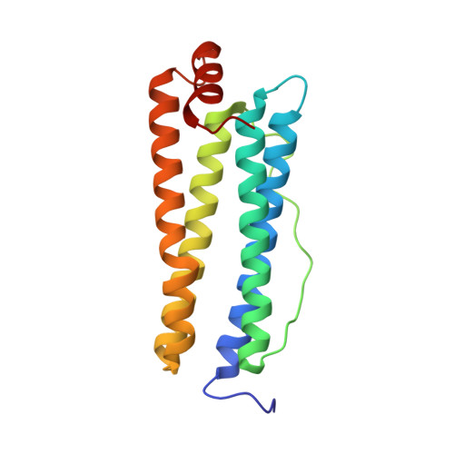
Preparation of modified and hybrid ferritin provides a great opportunity to understand the mechanisms of iron loading/unloading, protein self-assembly, size constrained nanomaterial synthesis and targeted drug delivery. However, the large size (M.W.=490kDa) has been limiting the separation of different modified and/or hybrid ferritin nanocages from each other in their intact assembled form and further characterization. Native polyacrylamide gel electrophoresis (PAGE) separates proteins on the basis of both charge and mass, while maintaining their overall native structure and activity. Altering surface charge distribution by substitution of amino acid residues located at the external surface of ferritin (K104E & D40A) affected the migration rate in native PAGE while internal modification had little effect. Crystal structures confirmed that ferritin nanocages made up of subunits with single amino acid substitutions retain the overall structure of ferritin nanocage. Taking advantage of K104E migration behavior, formation of hybrid ferritins with subunits of wild type (WT) and K104E were confirmed and separated in native PAGE. Cage integrity and iron loading ability (ferritin activity) were also tested. The migration pattern of hybrid ferritins in native PAGE depends on the subunit ratio (WT: K104E) in the ferritin cage. Our work shows that native PAGE can be exploited in nanobiotechnology, by analyzing modifications of large proteins like ferritin. Native PAGE, a simple, straight-forward technique, can be used to analyze small modification (by altering external surface charge) in large proteins like ferritin, without disintegrating its self-assembled nanocage structure. In doing so, native PAGE can complement the information obtained from mass spectrometry. The confirmation and separation of modified and hybrid ferritin protein nanocages in native PAGE, opens up various prospects of bio-conjugation, which can be useful in targeted drug delivery, nanobiotechnology and in understanding nature's idea of synthesizing hybrid ferritins in different human tissues.
- Department of Chemistry, National Institute of Technology, Rourkela 769008, Odisha, India; KIIT School of Biotechnology, KIIT University, Bhubaneswar 751024, Odisha, India.
Organizational Affiliation:


