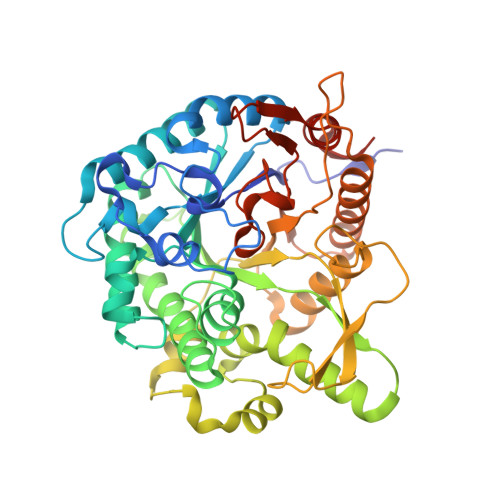
5XGZ

Metagenomic glucose-tolerant glycosidase


Experimental Data Snapshot

  • Method: X-RAY DIFFRACTION
  • Resolution: 1.75 Å
  • R-Value Free: 0.255 
  • R-Value Work: 0.206 
  • R-Value Observed: 0.209 

Starting Model: experimental
View more details

wwPDB Validation   3D Report Full Report


This is version 1.2 of the entry. See complete history


Literature

Improved thermostability of a metagenomic glucose-tolerant beta-glycosidase based on its X-ray crystal structure.

Matsuzawa, T.Watanabe, M.Yaoi, K.

(2017) Appl Microbiol Biotechnol 101: 8353-8363

  • DOI: https://doi.org/10.1007/s00253-017-8525-9
  • Primary Citation of Related Structures:  
    5XGZ

  • PubMed Abstract: 

    MeBglD2, a metagenomic β-glycosidase, is stimulated by various saccharides, including D-glucose, D-xylose, and maltose, and it promotes the enzymatic saccharification of plant biomass. To improve the thermostability of MeBglD2, its X-ray crystal structure was analyzed, and the amino acid residues responsible for its thermostability were identified using the structural information. Mutations in His8, Asn59, and Gly295 improved the thermostability of MeBglD2, and the combination of these mutations resulted in the highest thermostability. Compared with wild-type MeBglD2, thermostable MeBglD2 mutants promoted plant biomass saccharification using Trichoderma reesei cellulase. In addition to thermostability, the thermostable mutants exhibited higher tolerance to ethanol, dimethyl sulfoxide, and copper ions, indicating that the MeBglD2 mutants generated in this study were improved in their tolerance to not only high temperature but also to organic solvents and metal ions.


  • Organizational Affiliation

    Bioproduction Research Institute, National Institute of Advanced Industrial Science and Technology (AIST), Tsukuba Central 6, 1-1-1 Higashi, Tsukuba, Ibaraki, 305-8566, Japan. matsuzawa-tomohiko@aist.go.jp.


Macromolecules
Find similar proteins by:  (by identity cutoff)  |  3D Structure
Entity ID: 1
MoleculeChains Sequence LengthOrganismDetailsImage
Beta-glycosidase
A, B
457uncultured microorganismMutation(s): 0 
EC: 3.2.1.21
UniProt
Find proteins for A0A1E1FFN6 (uncultured microorganism)
Explore A0A1E1FFN6 
Go to UniProtKB:  A0A1E1FFN6
Entity Groups  
Sequence Clusters30% Identity50% Identity70% Identity90% Identity95% Identity100% Identity
UniProt GroupA0A1E1FFN6
Sequence Annotations
Expand
  • Reference Sequence
Small Molecules
Ligands 3 Unique
IDChains Name / Formula / InChI Key2D Diagram3D Interactions
SO4
Query on SO4

Download Ideal Coordinates CCD File 
L [auth A],
W [auth B]
SULFATE ION
O4 S
QAOWNCQODCNURD-UHFFFAOYSA-L
GOL
Query on GOL

Download Ideal Coordinates CCD File 
C [auth A]
D [auth A]
E [auth A]
F [auth A]
G [auth A]
C [auth A],
D [auth A],
E [auth A],
F [auth A],
G [auth A],
H [auth A],
I [auth A],
M [auth B],
N [auth B],
O [auth B],
P [auth B],
Q [auth B],
R [auth B],
S [auth B],
T [auth B]
GLYCEROL
C3 H8 O3
PEDCQBHIVMGVHV-UHFFFAOYSA-N
NI
Query on NI

Download Ideal Coordinates CCD File 
J [auth A],
K [auth A],
U [auth B],
V [auth B]
NICKEL (II) ION
Ni
VEQPNABPJHWNSG-UHFFFAOYSA-N
Experimental Data & Validation

Experimental Data

  • Method: X-RAY DIFFRACTION
  • Resolution: 1.75 Å
  • R-Value Free: 0.255 
  • R-Value Work: 0.206 
  • R-Value Observed: 0.209 
  • Space Group: I 2 3
Unit Cell:
Length ( Å )Angle ( ˚ )
a = 202.427α = 90
b = 202.427β = 90
c = 202.427γ = 90
Software Package:
Software NamePurpose
REFMACrefinement
DENZOdata reduction
SCALEPACKdata scaling
PHASERphasing

Structure Validation

View Full Validation Report


Entry History & Funding Information

Deposition Data


Funding OrganizationLocationGrant Number
JSPSJapan26850067

Revision History  (Full details and data files)

  • Version 1.0: 2018-05-02
    Type: Initial release
  • Version 1.1: 2019-11-13
    Changes: Database references
  • Version 1.2: 2023-11-22
    Changes: Data collection, Database references, Derived calculations, Refinement description