5XE8 | pdb_00005xe8

Crystal Structure of the Peptidase Domain of Streptococcus mutans ComA


Experimental Data Snapshot

  • Method: X-RAY DIFFRACTION
  • Resolution: 3.10 Å
  • R-Value Free: 
    0.269 (Depositor), 0.271 (DCC) 
  • R-Value Work: 
    0.236 (Depositor), 0.239 (DCC) 
  • R-Value Observed: 
    0.238 (Depositor) 

Starting Model: experimental
View more details

wwPDB Validation 3D Report Full Report

Validation slider image for 5XE8

This is version 1.3 of the entry. See complete history

Literature

High-throughput Screening of Small Molecule Inhibitors of the Streptococcus Quorum-sensing Signal Pathway

Ishii, S.Fukui, K.Yokoshima, S.Kumagai, K.Beniyama, Y.Kodama, T.Fukuyama, T.Okabe, T.Nagano, T.Kojima, H.Yano, T.

(2017) Sci Rep 7: 4029-4029

  • DOI: https://doi.org/10.1038/s41598-017-03567-2
  • Primary Citation Related Structures: 
    5XE8, 5XE9

  • PubMed Abstract: 

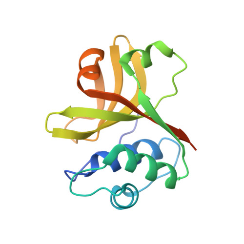
    The main components of the quorum-sensing system are expected to be favorable targets for drug development to combat various chronic infectious diseases. ComA of Streptococcus is an ATP-binding cassette transporter containing a peptidase domain (PEP), which is essential for the quorum-sensing signal production. Using high-throughput screening, we found a potent small molecule that suppressed the S. mutans quorum-sensing pathway through inhibition of PEP activity. The compound effectively attenuated the biofilm formation and competence development of S. mutans without inhibiting cell growth. The kinetic and structural studies with this molecule and a related compound unexpectedly revealed an allosteric site of PEP. This relatively hydrophobic site is thought to undergo large structural changes during the catalytic process. These compounds inhibit PEP activity by binding to and suppressing the structural changes of this site. These results showed that PEP is a good target for inhibitors of the Streptococcus quorum-sensing system.


  • Organizational Affiliation
    • Department of Biochemistry, Osaka Medical College, Takatsuki, Osaka, 569-8686, Japan. med002@osaka-med.ac.jp.

Macromolecule Content 

  • Total Structure Weight: 34.92 kDa 
  • Atom Count: 2,055 
  • Modeled Residue Count: 257 
  • Deposited Residue Count: 304 
  • Unique protein chains: 1

Macromolecules

Find similar proteins by:|  3D Structure
Entity ID: 1
MoleculeChains  Sequence LengthOrganismDetailsImage
Putative ABC transporter, ATP-binding protein ComAA,
B [auth C]
152Streptococcus mutans UA159Mutation(s): 0 
Gene Names: SMU_286
UniProt
Find proteins for Q8DW05 (Streptococcus mutans serotype c (strain ATCC 700610 / UA159))
Explore Q8DW05 
Go to UniProtKB:  Q8DW05
Entity Groups
Sequence Clusters30% Identity50% Identity70% Identity90% Identity95% Identity100% Identity
UniProt GroupQ8DW05
Sequence Annotations
Expand
Reference Sequence

Experimental Data & Validation

Experimental Data

  • Method: X-RAY DIFFRACTION
  • Resolution: 3.10 Å
  • R-Value Free:  0.269 (Depositor), 0.271 (DCC) 
  • R-Value Work:  0.236 (Depositor), 0.239 (DCC) 
  • R-Value Observed: 0.238 (Depositor) 
Space Group: P 21 21 21
Unit Cell:
Length ( Å )Angle ( ˚ )
a = 47.071α = 90
b = 59.437β = 90
c = 121.673γ = 90
Software Package:
Software NamePurpose
PHENIXrefinement
HKL-2000data scaling
PHENIXphasing
REFMACrefinement
PHENIXmodel building
HKL-2000data reduction

Structure Validation

View Full Validation Report


Entry History 

Deposition Data

Revision History  (Full details and data files)

  • Version 1.0: 2017-06-28
    Type: Initial release
  • Version 1.1: 2017-08-16
    Changes: Database references
  • Version 1.2: 2023-10-11
    Changes: Data collection, Database references, Refinement description
  • Version 1.3: 2023-11-22
    Changes: Refinement description