5XBJ | pdb_00005xbj

The structure of the flagellar hook junction protein HAP1 (FlgK) from Campylobacter jejuni


Experimental Data Snapshot

  • Method: X-RAY DIFFRACTION
  • Resolution: 2.45 Å
  • R-Value Free: 
    0.232 (Depositor), 0.229 (DCC) 
  • R-Value Work: 
    0.178 (Depositor), 0.179 (DCC) 
  • R-Value Observed: 
    0.183 (Depositor) 

Starting Model: experimental
View more details

wwPDB Validation 3D Report Full Report

Validation slider image for 5XBJ

This is version 1.1 of the entry. See complete history

Literature

Structure of FlgK reveals the divergence of the bacterial Hook-Filament Junction of Campylobacter

Bulieris, P.V.Shaikh, N.H.Freddolino, P.L.Samatey, F.A.

(2017) Sci Rep 7: 15743-15743

  • DOI: https://doi.org/10.1038/s41598-017-15837-0
  • Primary Citation Related Structures: 
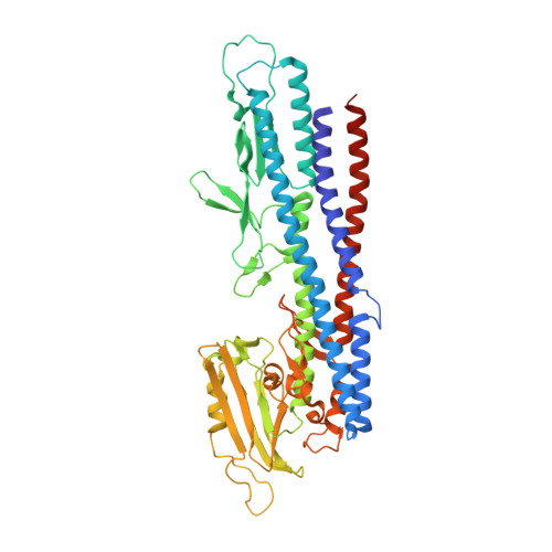
    5XBJ

  • PubMed Abstract: 

    Evolution of a nano-machine consisting of multiple parts, each with a specific function, is a complex process. A change in one part should eventually result in changes in other parts, if the overall function is to be conserved. In bacterial flagella, the filament and the hook have distinct functions and their respective proteins, FliC and FlgE, have different three-dimensional structures. The filament functions as a helical propeller and the hook as a flexible universal joint. Two proteins, FlgK and FlgL, assure a smooth connectivity between the hook and the filament. Here we show that, in Campylobacter, the 3D structure of FlgK differs from that of its orthologs in Salmonella and Burkholderia, whose structures have previously been solved. Docking the model of the FlgK junction onto the structure of the Campylobacter hook provides some clues about its divergence. These data suggest how evolutionary pressure to adapt to structural constraints, due to the structure of Campylobacter hook, causes divergence of one element of a supra-molecular complex in order to maintain the function of the entire flagellar assembly.


  • Organizational Affiliation
    • Trans-membrane Trafficking Unit, Okinawa Institute of Science and Technology Graduate University, 1919-1 Tancha, Onna, Kunigami, Okinawa, 904-0495, Japan.

Macromolecule Content 

  • Total Structure Weight: 56.98 kDa 
  • Atom Count: 4,322 
  • Modeled Residue Count: 500 
  • Deposited Residue Count: 511 
  • Unique protein chains: 1

Macromolecules

Find similar proteins by:|  3D Structure
Entity ID: 1
MoleculeChains  Sequence LengthOrganismDetailsImage
Flagellar hook-associated protein FlgK511Campylobacter jejuniMutation(s): 0 
UniProt
Find proteins for Q0P8E9 (Campylobacter jejuni subsp. jejuni serotype O:2 (strain ATCC 700819 / NCTC 11168))
Explore Q0P8E9 
Go to UniProtKB:  Q0P8E9
Entity Groups
Sequence Clusters30% Identity50% Identity70% Identity90% Identity95% Identity100% Identity
UniProt GroupQ0P8E9
Sequence Annotations
Expand
Reference Sequence

Experimental Data & Validation

Experimental Data

  • Method: X-RAY DIFFRACTION
  • Resolution: 2.45 Å
  • R-Value Free:  0.232 (Depositor), 0.229 (DCC) 
  • R-Value Work:  0.178 (Depositor), 0.179 (DCC) 
  • R-Value Observed: 0.183 (Depositor) 
Space Group: P 21 21 2
Unit Cell:
Length ( Å )Angle ( ˚ )
a = 49.501α = 90
b = 123.552β = 90
c = 92.54γ = 90
Software Package:
Software NamePurpose
PHENIXrefinement
HKL-2000data reduction
HKL-2000data scaling
PHASERphasing

Structure Validation

View Full Validation Report


Entry History 

& Funding Information

Deposition Data


Funding OrganizationLocationGrant Number
Okinawa Institute of Science and Technology Graduate UniversityJapan--

Revision History  (Full details and data files)

  • Version 1.0: 2017-12-06
    Type: Initial release
  • Version 1.1: 2023-11-22
    Changes: Data collection, Database references, Refinement description