5X4A

SLL-2-Forssman antigen tetrasaccharides complex


Experimental Data Snapshot

  • Method: X-RAY DIFFRACTION
  • Resolution: 3.40 Å
  • R-Value Free: 0.266 
  • R-Value Work: 0.239 

Starting Model: experimental
View more details

wwPDB Validation   3D Report Full Report


Ligand Structure Quality Assessment 


This is version 2.1 of the entry. See complete history


Literature

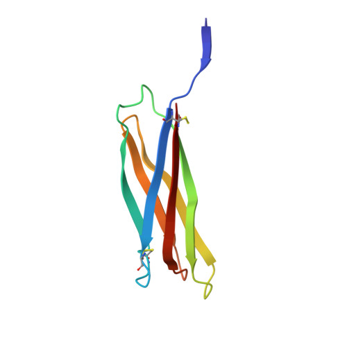
Crystal structure of octocoral lectin SLL-2 complexed with Forssman antigen tetrasaccharide.

Kita, A.Jimbo, M.Sakai, R.Morimoto, Y.Takeuchi, R.Tanaka, H.Takahashi, T.Miki, K.

(2017) Glycobiology 

  • DOI: https://doi.org/10.1093/glycob/cwx043
  • Primary Citation of Related Structures:  
    5X4A

  • PubMed Abstract: 

    A symbiosis-related lectin, SLL-2, from the octocoral Sinularia lochmodes, distributes densely on the cell surface of microalgae, Symbiodinium sp., an endosymbiotic dinoflagellate of the coral, and is also shown to be a chemical cue that transforms dinoflagellates into a nonmotile (coccoid) symbiotic state. SLL-2 binds to the sugar chain of the molecule similar to Forssman antigen pentasaccharide (GalNAcα1-3GalNAcβ1-3 Galα1-4 Galβ1-4Glc) on the surface of microalgae with high affinity. Here we report the crystal structure of the complex between SLL-2 and Forssman antigen tetrasaccharide (GalNAcα1-3GalNAcβ1-3 Galα1-4 Galβ) at 3.4 Å resolution. In an asymmetric unit of the crystal, there are two hexameric molecules with totally 12 sugar recognition sites. At 9 in 12 sites, the first and second saccharides of the Forssman antigen tetrasaccharide bind directly to galactopyranoside binding site of SLL-2, whereas the third and fourth saccharides have no interaction with the SLL-2 hexameric molecule that binds the first saccharide. The sugar chain bends at α-1,4-glycosidic linkage between the third and fourth saccharides toward the position that we defined as a pyranoside binding site in the crystal structure of the complex between SLL-2 and GalNAc. The structure allowed us to suggest a possible binding mode of the Forssman antigen pentasaccharide to SLL-2. These observations support our hypothesis that the binding of SLL-2 to the cell surface sugars of zooxanthella in a unique manner might trigger some physiological changes of the cell to adapt symbiosis with the host coral.


  • Organizational Affiliation

    Research Reactor Institute, Kyoto University, Kumatori, Sennan, Osaka 590-0494, Japan.


Macromolecules
Find similar proteins by:  (by identity cutoff)  |  3D Structure
Entity ID: 1
MoleculeChains Sequence LengthOrganismDetailsImage
Galactose-binding lectin
A, B, C, D, E
A, B, C, D, E, F, G, H, I, J, K, L
94Sinularia lochmodesMutation(s): 0 
UniProt
Find proteins for A4CYJ6 (Sinularia lochmodes)
Explore A4CYJ6 
Go to UniProtKB:  A4CYJ6
Entity Groups  
Sequence Clusters30% Identity50% Identity70% Identity90% Identity95% Identity100% Identity
UniProt GroupA4CYJ6
Glycosylation
Glycosylation Sites: 1
Sequence Annotations
Expand
  • Reference Sequence
Oligosaccharides

Help

Entity ID: 2
MoleculeChains Length2D Diagram Glycosylation3D Interactions
2-acetamido-2-deoxy-beta-D-glucopyranose-(1-4)-2-acetamido-2-deoxy-beta-D-glucopyranose
BA [auth b],
CA [auth c],
EA [auth e],
GA [auth g],
M,
BA [auth b],
CA [auth c],
EA [auth e],
GA [auth g],
M,
O,
Q,
R,
T,
V,
X,
Z
2N-Glycosylation
Glycosylation Resources
GlyTouCan:  G42666HT
GlyCosmos:  G42666HT
GlyGen:  G42666HT
Entity ID: 3
MoleculeChains Length2D Diagram Glycosylation3D Interactions
2-acetamido-2-deoxy-alpha-D-galactopyranose-(1-3)-2-acetamido-2-deoxy-beta-D-galactopyranose-(1-3)-alpha-D-galactopyranose-(1-4)-beta-D-galactopyranose
AA [auth a],
DA [auth d],
FA [auth f],
HA [auth h],
N,
AA [auth a],
DA [auth d],
FA [auth f],
HA [auth h],
N,
P,
S,
U,
W,
Y
4N/A
Glycosylation Resources
GlyTouCan:  G88796WA
GlyCosmos:  G88796WA
Small Molecules
Ligands 1 Unique
IDChains Name / Formula / InChI Key2D Diagram3D Interactions
GLA
Query on GLA

Download Ideal Coordinates CCD File 
IA [auth A]alpha-D-galactopyranose
C6 H12 O6
WQZGKKKJIJFFOK-PHYPRBDBSA-N
Experimental Data & Validation

Experimental Data

  • Method: X-RAY DIFFRACTION
  • Resolution: 3.40 Å
  • R-Value Free: 0.266 
  • R-Value Work: 0.239 
  • Space Group: P 21 21 21
Unit Cell:
Length ( Å )Angle ( ˚ )
a = 96.468α = 90
b = 99.939β = 90
c = 273.936γ = 90
Software Package:
Software NamePurpose
CNSrefinement
HKL-2000data reduction
HKL-2000data scaling
MOLREPphasing

Structure Validation

View Full Validation Report


Ligand Structure Quality Assessment 


Entry History & Funding Information

Deposition Data


Funding OrganizationLocationGrant Number
JSPSJapan15K12750

Revision History  (Full details and data files)

  • Version 1.0: 2017-09-20
    Type: Initial release
  • Version 2.0: 2020-07-29
    Type: Remediation
    Reason: Carbohydrate remediation
    Changes: Advisory, Atomic model, Data collection, Derived calculations, Structure summary
  • Version 2.1: 2023-11-22
    Changes: Data collection, Database references, Derived calculations, Refinement description, Structure summary