5WT9 | pdb_00005wt9

Complex structure of PD-1 and nivolumab-Fab


Experimental Data Snapshot

  • Method: X-RAY DIFFRACTION
  • Resolution: 2.40 Å
  • R-Value Free: 
    0.228 (Depositor), 0.230 (DCC) 
  • R-Value Work: 
    0.187 (Depositor), 0.189 (DCC) 
  • R-Value Observed: 
    0.189 (Depositor) 

Starting Model: experimental
View more details

wwPDB Validation 3D Report Full Report

Validation slider image for 5WT9

This is version 2.2 of the entry. See complete history

Literature

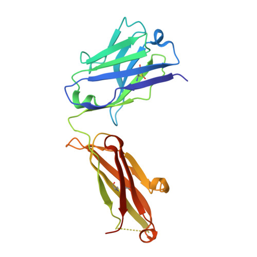
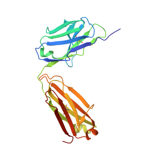
An unexpected N-terminal loop in PD-1 dominates binding by nivolumab.

Tan, S.Zhang, H.Chai, Y.Song, H.Tong, Z.Wang, Q.Qi, J.Wong, G.Zhu, X.Liu, W.J.Gao, S.Wang, Z.Shi, Y.Yang, F.Gao, G.F.Yan, J.

(2017) Nat Commun 8: 14369-14369

  • DOI: https://doi.org/10.1038/ncomms14369
  • Primary Citation Related Structures: 
    5WT9

  • PubMed Abstract: 

    Cancer immunotherapy by targeting of immune checkpoint molecules has been a research 'hot-spot' in recent years. Nivolumab, a human monoclonal antibody targeting PD-1, has been widely used clinically since 2014. However, the binding mechanism of nivolumab to PD-1 has not yet been shown, despite a recent report describing the complex structure of pembrolizumab/PD-1. It has previously been speculated that PD-1 glycosylation is involved in nivolumab recognition. Here we report the complex structure of nivolumab with PD-1 and evaluate the effects of PD-1 N-glycosylation on the interactions with nivolumab. Structural and functional analyses unexpectedly reveal an N-terminal loop outside the IgV domain of PD-1. This loop is not involved in recognition of PD-L1 but dominates binding to nivolumab, whereas N-glycosylation is not involved in binding at all. Nivolumab binds to a completely different area than pembrolizumab. These results provide the basis for the design of future inhibitory molecules targeting PD-1.


  • Organizational Affiliation
    • CAS Key Laboratory of Microbial Physiological and Metabolic engineering, Institute of Microbiology, Chinese Academy of Sciences, Beijing 100101, China.

Macromolecule Content 

  • Total Structure Weight: 67.54 kDa 
  • Atom Count: 4,391 
  • Modeled Residue Count: 533 
  • Deposited Residue Count: 610 
  • Unique protein chains: 3

Macromolecules

Find similar proteins by:|  3D Structure
Entity ID: 1
MoleculeChains  Sequence LengthOrganismDetailsImage
Heavy Chain of NivolumabA [auth H]220Homo sapiensMutation(s): 0 
Find similar proteins by:|  3D Structure
Entity ID: 2
MoleculeChains  Sequence LengthOrganismDetailsImage
Light Chain of NivolumabB [auth L]217Homo sapiensMutation(s): 0 
Find similar proteins by:|  3D Structure
Entity ID: 3
MoleculeChains  Sequence LengthOrganismDetailsImage
Programmed cell death protein 1C [auth G]173Homo sapiensMutation(s): 0 
Gene Names: PDCD1PD1
UniProt & NIH Common Fund Data Resources
Find proteins for Q15116 (Homo sapiens)
Explore Q15116 
Go to UniProtKB:  Q15116
PHAROS:  Q15116
GTEx:  ENSG00000188389 
Entity Groups
Sequence Clusters30% Identity50% Identity70% Identity90% Identity95% Identity100% Identity
UniProt GroupQ15116
Glycosylation
Glycosylation Sites: 1Go to GlyGen: Q15116-1
Sequence Annotations
Expand
Reference Sequence

Oligosaccharides

Help  
Entity ID: 4
MoleculeChains Length2D Diagram GlycosylationD Interactions
2-acetamido-2-deoxy-beta-D-glucopyranose-(1-4)-[alpha-L-fucopyranose-(1-6)]2-acetamido-2-deoxy-beta-D-glucopyranoseD [auth A]3N-Glycosylation
Glycosylation Resources
GlyTouCan: G21290RB
GlyCosmos: G21290RB
GlyGen: G21290RB

Experimental Data & Validation

Experimental Data

  • Method: X-RAY DIFFRACTION
  • Resolution: 2.40 Å
  • R-Value Free:  0.228 (Depositor), 0.230 (DCC) 
  • R-Value Work:  0.187 (Depositor), 0.189 (DCC) 
  • R-Value Observed: 0.189 (Depositor) 
Space Group: P 43 21 2
Unit Cell:
Length ( Å )Angle ( ˚ )
a = 108.527α = 90
b = 108.527β = 90
c = 145.423γ = 90
Software Package:
Software NamePurpose
PHENIXrefinement
HKL-2000data reduction
HKL-2000data scaling
PHASERphasing

Structure Validation

View Full Validation Report


Entry History 

& Funding Information

Deposition Data


Funding OrganizationLocationGrant Number
Scientific Research and Development of ChinaChina2016YFC1200300
National Natural Science Foundation of ChinaChina31390432
National Natural Science Foundation of ChinaChina31500722
Chinese Academy of SciencesChina153211KYSB20160001
China National Grand S&T Special ProjectChina2013ZX10004608-002
National Natural Science Foundation of ChinaChina81321063

Revision History  (Full details and data files)

  • Version 1.0: 2017-02-15
    Type: Initial release
  • Version 1.1: 2017-12-13
    Changes: Database references
  • Version 2.0: 2020-07-29
    Type: Remediation
    Reason: Carbohydrate remediation
    Changes: Atomic model, Data collection, Derived calculations, Structure summary
  • Version 2.1: 2023-11-08
    Changes: Data collection, Database references, Refinement description, Structure summary
  • Version 2.2: 2024-10-23
    Changes: Structure summary