5WRU | pdb_00005wru

Crystal structure of type I inorganic pyrophosphatase from P falciparum


Experimental Data Snapshot

  • Method: X-RAY DIFFRACTION
  • Resolution: 3.19 Å
  • R-Value Free: 
    0.277 (Depositor), 0.267 (DCC) 
  • R-Value Work: 
    0.234 (Depositor), 0.233 (DCC) 
  • R-Value Observed: 
    0.237 (Depositor) 

wwPDB Validation 3D Report Full Report

Validation slider image for 5WRU

This is version 1.2 of the entry. See complete history

Literature

Structural and Biochemical Characterization of Apicomplexan Inorganic Pyrophosphatases

Jamwal, A.Yogavel, M.Abdin, M.Z.Jain, S.K.Sharma, A.

(2017) Sci Rep 7: 5255-5255

  • DOI: https://doi.org/10.1038/s41598-017-05234-y
  • Primary Citation Related Structures: 
    5WRT, 5WRU

  • PubMed Abstract: 

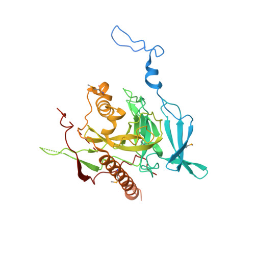
    Inorganic pyrophosphatases (PPase) participate in energy cycling and they are essential for growth and survival of organisms. Here we report extensive structural and functional characterization of soluble PPases from the human parasites Plasmodium falciparum (PfPPase) and Toxoplasma gondii (TgPPase). Our results show that PfPPase is a cytosolic enzyme whose gene expression is upregulated during parasite asexual stages. Cambialistic PfPPase actively hydrolyzes linear short chain polyphosphates like PP i , polyP 3 and ATP in the presence of Zn 2+ . A remarkable new feature of PfPPase is the low complexity asparagine-rich N-terminal region that mediates its dimerization. Deletion of N-region has an unexpected and substantial effect on the stability of PfPPase domain, resulting in aggregation and significant loss of enzyme activity. Significantly, the crystal structures of PfPPase and TgPPase reveal unusual and unprecedented dimeric organizations and provide new fundamental insights into the variety of oligomeric assemblies possible in eukaryotic inorganic PPases.


  • Organizational Affiliation
    • Molecular Medicine Group, International Centre for Genetic Engineering and Biotechnology, Aruna Asaf Ali Marg, New Delhi, 110067, India.

Macromolecule Content 

  • Total Structure Weight: 228.06 kDa 
  • Atom Count: 13,039 
  • Modeled Residue Count: 1,543 
  • Deposited Residue Count: 1,900 
  • Unique protein chains: 1

Macromolecules

Find similar proteins by:|  3D Structure
Entity ID: 1
MoleculeChains  Sequence LengthOrganismDetailsImage
Probable inorganic pyrophosphatase
A, B, C, D, E
380Plasmodium falciparum 3D7Mutation(s): 0 
Gene Names: MAL3P6.3PFC0710w
EC: 3.6.1.1
UniProt
Find proteins for O77392 (Plasmodium falciparum (isolate 3D7))
Explore O77392 
Go to UniProtKB:  O77392
Entity Groups
Sequence Clusters30% Identity50% Identity70% Identity90% Identity95% Identity100% Identity
UniProt GroupO77392
Sequence Annotations
Expand
Reference Sequence

Small Molecules

Modified Residues  1 Unique
IDChains TypeFormula2D DiagramParent
MSE
Query on MSE
A, B, C, D, E
L-PEPTIDE LINKINGC5 H11 N O2 SeMET

Experimental Data & Validation

Experimental Data

  • Method: X-RAY DIFFRACTION
  • Resolution: 3.19 Å
  • R-Value Free:  0.277 (Depositor), 0.267 (DCC) 
  • R-Value Work:  0.234 (Depositor), 0.233 (DCC) 
  • R-Value Observed: 0.237 (Depositor) 
Space Group: C 1 2 1
Unit Cell:
Length ( Å )Angle ( ˚ )
a = 253.345α = 90
b = 85.22β = 114.95
c = 108.445γ = 90
Software Package:
Software NamePurpose
PDB_EXTRACTdata extraction
HKL-2000data reduction
HKL-2000data scaling
AutoSolphasing
PHENIXrefinement

Structure Validation

View Full Validation Report


Entry History 

Deposition Data

Revision History  (Full details and data files)

  • Version 1.0: 2017-10-11
    Type: Initial release
  • Version 1.1: 2025-04-09
    Changes: Data collection, Database references, Refinement description, Structure summary
  • Version 1.2: 2025-09-17
    Changes: Advisory, Derived calculations, Structure summary