5WR0 | pdb_00005wr0

Huisgen cycloaddition for PPARg-LBD labeling by soaking method


Experimental Data Snapshot

  • Method: X-RAY DIFFRACTION
  • Resolution: 2.85 Å
  • R-Value Free: 
    0.300 (Depositor), 0.297 (DCC) 
  • R-Value Work: 
    0.250 (Depositor), 0.249 (DCC) 
  • R-Value Observed: 
    0.252 (Depositor) 

wwPDB Validation 3D Report Full Report

Validation slider image for 5WR0

Ligand Structure Quality Assessment 


This is version 1.2 of the entry. See complete history

Literature

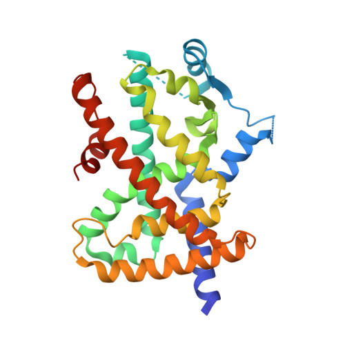
On-site reaction for PPAR gamma modification using a specific bifunctional ligand

Kojima, H.Itoh, T.Yamamoto, K.

(2017) Bioorg Med Chem 25: 6492-6500

  • DOI: https://doi.org/10.1016/j.bmc.2017.10.024
  • Primary Citation Related Structures: 
    5WQX, 5WR0, 5WR1

  • PubMed Abstract: 

    Site-specific labeling is an important methodology to elucidate the biological function of a target protein. Here, we report a strategy for site-specific chemical labeling, termed the "on-site reaction". We designed and readily synthesized a bifunctional ligand possessing two reaction sites, an enone and an azide moiety. This strategy involves an on-site conjugate addition reaction with protein followed by a Hüisgen cycloaddition reaction. We demonstrate this strategy by using fluorescein as a probe and peroxisome proliferator activated receptor γ (PPARγ) as a target protein. The reactions were evaluated by ESI-mass analysis and the binding site and modes of binding were revealed by X-ray crystallization analysis. The proposed methodology can easily convert a covalent ligand into chemical tool for protein functional analysis and the identification of drug targets.


  • Organizational Affiliation
    • Laboratory of Drug Design and Medicinal Chemistry, Showa Pharmaceutical University, 3-3165 Higashi-Tamagawagakuen, Machida, Tokyo 194-8543, Japan.

Macromolecule Content 

  • Total Structure Weight: 63.66 kDa 
  • Atom Count: 4,071 
  • Modeled Residue Count: 503 
  • Deposited Residue Count: 552 
  • Unique protein chains: 1

Macromolecules

Find similar proteins by:|  3D Structure
Entity ID: 1
MoleculeChains  Sequence LengthOrganismDetailsImage
Peroxisome proliferator-activated receptor gamma
A, B
276Homo sapiensMutation(s): 0 
Gene Names: PPARGNR1C3
UniProt & NIH Common Fund Data Resources
Find proteins for P37231 (Homo sapiens)
Explore P37231 
Go to UniProtKB:  P37231
PHAROS:  P37231
GTEx:  ENSG00000132170 
Entity Groups
Sequence Clusters30% Identity50% Identity70% Identity90% Identity95% Identity100% Identity
UniProt GroupP37231
Sequence Annotations
Expand
Reference Sequence

Small Molecules

Ligands 1 Unique
IDChains Name / Formula / InChI Key2D Diagram3D Interactions
8Y0

Query on 8Y0


Download:Ideal Coordinates CCD File
C [auth A](E)-N-[(3E)-2-oxo-16-(8-{6-[(trifluoroacetyl)amino]hexanoyl}-8,9-dihydro-1H-dibenzo[b,f][1,2,3]triazolo[4,5-d]azocin-1-yl)hexadec-3-en-1-ylidene]glycine
C41 H51 F3 N6 O5
XTXBLKQONYTAIB-ABCDOUFNSA-N

Experimental Data & Validation

Experimental Data

  • Method: X-RAY DIFFRACTION
  • Resolution: 2.85 Å
  • R-Value Free:  0.300 (Depositor), 0.297 (DCC) 
  • R-Value Work:  0.250 (Depositor), 0.249 (DCC) 
  • R-Value Observed: 0.252 (Depositor) 
Space Group: C 1 2 1
Unit Cell:
Length ( Å )Angle ( ˚ )
a = 92.73α = 90
b = 61.21β = 102.95
c = 118.35γ = 90
Software Package:
Software NamePurpose
REFMACrefinement

Structure Validation

View Full Validation Report


Ligand Structure Quality Assessment 


Entry History 

Deposition Data

Revision History  (Full details and data files)

  • Version 1.0: 2017-11-22
    Type: Initial release
  • Version 1.1: 2017-12-13
    Changes: Database references
  • Version 1.2: 2024-11-20
    Changes: Data collection, Database references, Structure summary