5WO2

Chaperone Spy bound to Casein Fragment (Casein un-modeled)


Experimental Data Snapshot

  • Method: X-RAY DIFFRACTION
  • Resolution: 1.77 Å
  • R-Value Free: 0.252 
  • R-Value Work: 0.215 
  • R-Value Observed: 0.216 

wwPDB Validation   3D Report Full Report


This is version 1.1 of the entry. See complete history

Re-refinement Note

This entry reflects an alternative modeling of the original data in: 5IOG


Literature

Visualizing chaperone-assisted protein folding.

Horowitz, S.Salmon, L.Koldewey, P.Ahlstrom, L.S.Martin, R.Quan, S.Afonine, P.V.van den Bedem, H.Wang, L.Xu, Q.Trievel, R.C.Brooks, C.L.Bardwell, J.C.

(2016) Nat Struct Mol Biol 23: 691-697

  • DOI: https://doi.org/10.1038/nsmb.3237
  • Primary Citation of Related Structures:  
    5WNW, 5WO1, 5WO2, 5WO3

  • PubMed Abstract: 

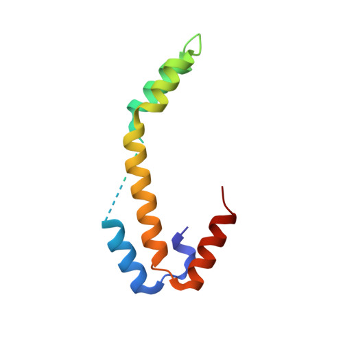
    Challenges in determining the structures of heterogeneous and dynamic protein complexes have greatly hampered past efforts to obtain a mechanistic understanding of many important biological processes. One such process is chaperone-assisted protein folding. Obtaining structural ensembles of chaperone-substrate complexes would ultimately reveal how chaperones help proteins fold into their native state. To address this problem, we devised a new structural biology approach based on X-ray crystallography, termed residual electron and anomalous density (READ). READ enabled us to visualize even sparsely populated conformations of the substrate protein immunity protein 7 (Im7) in complex with the Escherichia coli chaperone Spy, and to capture a series of snapshots depicting the various folding states of Im7 bound to Spy. The ensemble shows that Spy-associated Im7 samples conformations ranging from unfolded to partially folded to native-like states and reveals how a substrate can explore its folding landscape while being bound to a chaperone.


  • Organizational Affiliation

    Department of Molecular, Cellular, and Developmental Biology, University of Michigan, Ann Arbor, Michigan, USA.


Macromolecules
Find similar proteins by:  (by identity cutoff)  |  3D Structure
Entity ID: 1
MoleculeChains Sequence LengthOrganismDetailsImage
Periplasmic chaperone Spy
A, B
97Escherichia coliMutation(s): 0 
UniProt
Find proteins for P77754 (Escherichia coli (strain K12))
Explore P77754 
Go to UniProtKB:  P77754
Entity Groups  
Sequence Clusters30% Identity50% Identity70% Identity90% Identity95% Identity100% Identity
UniProt GroupP77754
Sequence Annotations
Expand
  • Reference Sequence
Small Molecules
Ligands 3 Unique
IDChains Name / Formula / InChI Key2D Diagram3D Interactions
IMD
Query on IMD

Download Ideal Coordinates CCD File 
C [auth A]
D [auth A]
Q [auth B]
S [auth B]
T [auth B]
C [auth A],
D [auth A],
Q [auth B],
S [auth B],
T [auth B],
U [auth B]
IMIDAZOLE
C3 H5 N2
RAXXELZNTBOGNW-UHFFFAOYSA-O
ZN
Query on ZN

Download Ideal Coordinates CCD File 
E [auth A]
F [auth A]
G [auth A]
H [auth A]
I [auth A]
E [auth A],
F [auth A],
G [auth A],
H [auth A],
I [auth A],
J [auth A],
L [auth A],
M [auth A],
N [auth A],
O [auth A],
R [auth B],
V [auth B],
W [auth B],
X [auth B],
Y [auth B],
Z [auth B]
ZINC ION
Zn
PTFCDOFLOPIGGS-UHFFFAOYSA-N
CL
Query on CL

Download Ideal Coordinates CCD File 
K [auth A],
P [auth A]
CHLORIDE ION
Cl
VEXZGXHMUGYJMC-UHFFFAOYSA-M
Experimental Data & Validation

Experimental Data

  • Method: X-RAY DIFFRACTION
  • Resolution: 1.77 Å
  • R-Value Free: 0.252 
  • R-Value Work: 0.215 
  • R-Value Observed: 0.216 
  • Space Group: P 41 2 2
Unit Cell:
Length ( Å )Angle ( ˚ )
a = 42.96α = 90
b = 42.96β = 90
c = 258.169γ = 90
Software Package:
Software NamePurpose
PHENIXrefinement
iMOSFLMdata reduction
Aimlessdata scaling
PHENIXphasing

Structure Validation

View Full Validation Report


Entry History 

Deposition Data

Revision History  (Full details and data files)

  • Version 1.0: 2017-08-16
    Type: Initial release
  • Version 1.1: 2023-10-04
    Changes: Data collection, Database references, Derived calculations, Refinement description