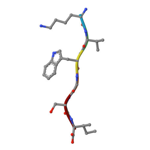
5WMJ

KVWGSI segment from Superoxide Dismutase 1,residues 30-35

  • Classification: PROTEIN FIBRIL
  • Organism(s): Homo sapiens
  • Mutation(s): No 

  • Deposited: 2017-07-28 Released: 2018-03-28 
  • Deposition Author(s): Sangwan, S., Sawaya, M., Eisenberg, D.
  • Funding Organization(s): National Institutes of Health/National Institute on Aging (NIH/NIA), Howard Hughes Medical Institute (HHMI)

Experimental Data Snapshot

  • Method: X-RAY DIFFRACTION
  • Resolution: 1.40 Å
  • R-Value Free: 0.170 
  • R-Value Work: 0.171 
  • R-Value Observed: 0.171 

Starting Model: in silico
View more details

wwPDB Validation   3D Report Full Report


This is version 1.5 of the entry. See complete history


Literature

Atomic structures of corkscrew-forming segments of SOD1 reveal varied oligomer conformations.

Sangwan, S.Sawaya, M.R.Murray, K.A.Hughes, M.P.Eisenberg, D.S.

(2018) Protein Sci 27: 1231-1242

  • DOI: https://doi.org/10.1002/pro.3391
  • Primary Citation of Related Structures:  
    5WMJ, 5WOR, 6B79

  • PubMed Abstract: 

    The aggregation cascade of disease-related amyloidogenic proteins, terminating in insoluble amyloid fibrils, involves intermediate oligomeric states. The structural and biochemical details of these oligomers have been largely unknown. Here we report crystal structures of variants of the cytotoxic oligomer-forming segment residues 28-38 of the ALS-linked protein, SOD1. The crystal structures reveal three different architectures: corkscrew oligomeric structure, nontwisting curved sheet structure and a steric zipper proto-filament structure. Our work highlights the polymorphism of the segment 28-38 of SOD1 and identifies the molecular features of amyloidogenic entities.


  • Organizational Affiliation

    Department of Biological Chemistry Los Angeles, Howard Hughes Medical Institute, UCLA-DOE and Molecular Biology Institute, California.


Macromolecules

Find similar proteins by:  Sequence   |   3D Structure  

Entity ID: 1
MoleculeChains Sequence LengthOrganismDetailsImage
Superoxide dismutase [Cu-Zn]
A, B
6Homo sapiensMutation(s): 0 
EC: 1.15.1.1
UniProt & NIH Common Fund Data Resources
Find proteins for P00441 (Homo sapiens)
Explore P00441 
Go to UniProtKB:  P00441
PHAROS:  P00441
GTEx:  ENSG00000142168 
Entity Groups  
UniProt GroupP00441
Sequence Annotations
Expand
  • Reference Sequence
Small Molecules
Ligands 1 Unique
IDChains Name / Formula / InChI Key2D Diagram3D Interactions
TFA
Query on TFA

Download Ideal Coordinates CCD File 
C [auth B]trifluoroacetic acid
C2 H F3 O2
DTQVDTLACAAQTR-UHFFFAOYSA-N
Experimental Data & Validation

Experimental Data

  • Method: X-RAY DIFFRACTION
  • Resolution: 1.40 Å
  • R-Value Free: 0.170 
  • R-Value Work: 0.171 
  • R-Value Observed: 0.171 
  • Space Group: P 21 21 21
Unit Cell:
Length ( Å )Angle ( ˚ )
a = 9.518α = 90
b = 20.282β = 90
c = 44.249γ = 90
Software Package:
Software NamePurpose
REFMACrefinement
DENZOdata reduction
SCALEPACKdata scaling
PHASERphasing

Structure Validation

View Full Validation Report


Entry History & Funding Information

Deposition Data


Funding OrganizationLocationGrant Number
National Institutes of Health/National Institute on Aging (NIH/NIA)United StatesAG054022
Howard Hughes Medical Institute (HHMI)United States--

Revision History  (Full details and data files)

  • Version 1.0: 2018-03-28
    Type: Initial release
  • Version 1.1: 2018-07-18
    Changes: Data collection, Database references
  • Version 1.2: 2019-11-06
    Changes: Author supporting evidence, Data collection
  • Version 1.3: 2019-11-20
    Changes: Author supporting evidence
  • Version 1.4: 2024-03-13
    Changes: Data collection, Database references
  • Version 1.5: 2024-04-03
    Changes: Refinement description