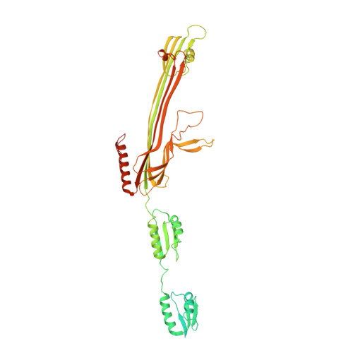
Structural Basis of Type 2 Secretion System Engagement between the Inner and Outer Bacterial Membranes.
Hay, I.D., Belousoff, M.J., Lithgow, T.(2017) mBio 8
- PubMed: 29042496
- DOI: https://doi.org/10.1128/mBio.01344-17
- Primary Citation of Related Structures:
5WLN - PubMed Abstract:
Sophisticated nanomachines are used by bacteria for protein secretion. In Gram-negative bacteria, the type 2 secretion system (T2SS) is composed of a pseudopilus assembly platform in the inner membrane and a secretin complex in the outer membrane. The engagement of these two megadalton-sized complexes is required in order to secrete toxins, effectors, and hydrolytic enzymes. Pseudomonas aeruginosa has at least two T2SSs, with the ancestral nanomachine having a secretin complex composed of XcpQ. Until now, no high-resolution structural information was available to distinguish the features of this Pseudomonas -type secretin, which varies greatly in sequence from the well-characterized Klebsiella -type and Vibrio -type secretins. We have purified the ~1-MDa secretin complex and analyzed it by cryo-electron microscopy. Structural comparisons with the Klebsiella -type secretin complex revealed a striking structural homology despite the differences in their sequence characteristics. At 3.6-Å resolution, the secretin complex was found to have 15-fold symmetry throughout the membrane-embedded region and through most of the domains in the periplasm. However, the N1 domain and N0 domain were not well ordered into this 15-fold symmetry. We suggest a model wherein this disordering of the subunit symmetry for the periplasmic N domains provides a means to engage with the 6-fold symmetry in the inner membrane platform, with a metastable engagement that can be disrupted by substrate proteins binding to the region between XcpP, in the assembly platform, and the XcpQ secretin. IMPORTANCE How the outer membrane and inner membrane components of the T2SS engage each other and yet can allow for substrate uptake into the secretin chamber has challenged the protein transport field for some time. This vexing question is of significance because the T2SS collects folded protein substrates in the periplasm for transport out of the bacterium and yet must discriminate these few substrate proteins from all the other hundred or so folded proteins in the periplasm. The structural analysis here supports a model wherein substrates must compete against a metastable interaction between XcpP in the assembly platform and the XcpQ secretin, wherein only structurally encoded features in the T2SS substrates compete well enough to disrupt XcpQ-XcpP for entry into the XcpQ chamber, for secretion across the outer membrane.
- Infection and Immunity Program, Biomedicine Discovery Institute and Department of Microbiology, Monash University, Clayton, Australia.
Organizational Affiliation:


