5WJB

Crystal Structure of Amino Acids 1733-1797 of Human Beta Cardiac Myosin Fused to Gp7


Experimental Data Snapshot

  • Method: X-RAY DIFFRACTION
  • Resolution: 2.90 Å
  • R-Value Free: 0.300 
  • R-Value Work: 0.249 
  • R-Value Observed: 0.251 

wwPDB Validation   3D Report Full Report


This is version 1.4 of the entry. See complete history


Literature

Design considerations in coiled-coil fusion constructs for the structural determination of a problematic region of the human cardiac myosin rod.

Andreas, M.P.Ajay, G.Gellings, J.A.Rayment, I.

(2017) J Struct Biol 200: 219-228

  • DOI: https://doi.org/10.1016/j.jsb.2017.07.006
  • Primary Citation of Related Structures:  
    5WJ7, 5WJB, 5WLQ, 5WLZ, 5WME

  • PubMed Abstract: 

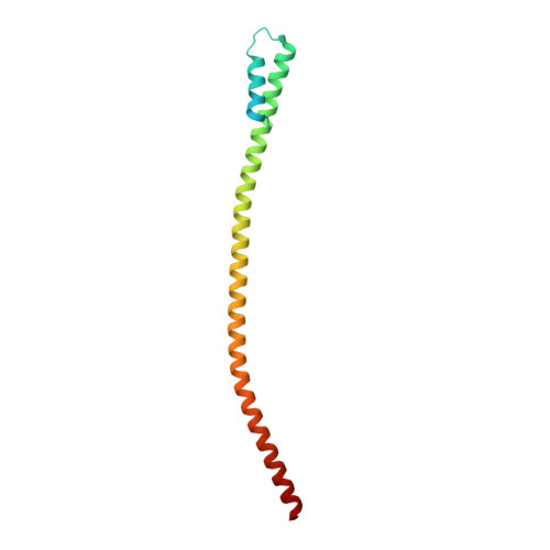
    X-ray structural determination of segments of the myosin rod has proved difficult because of the strong salt-dependent aggregation properties and repeating pattern of charges on the surface of the coiled-coil that lead to the formation of paracrystals. This problem has been resolved in part through the use of globular assembly domains that improve protein folding and prevent aggregation. The primary consideration now in designing coiled-coil fusion constructs for myosin is deciding where to truncate the coiled-coil and which amino acid residues to include from the folding domain. This is especially important for myosin that contains numerous regions of low predicted coiled-coil propensity. Here we describe the strategy adopted to determine the structure of the region that extends from Arg1677 - Leu1797 that included two areas that do not show a strong sequence signature of a conventional left-handed coiled coil or canonical heptad repeat. This demonstrates again that, with careful choice of fusion constructs, overlapping structures exhibit very similar conformations for the myosin rod fragments in the canonical regions. However, conformational variability is seen around Leu1706 which is a hot spot for cardiomyopathy mutations suggesting that this might be important for function.


  • Organizational Affiliation

    Department of Biochemistry, University of Wisconsin-Madison, WI 53706, USA.


Macromolecules
Find similar proteins by:  (by identity cutoff)  |  3D Structure
Entity ID: 1
MoleculeChains Sequence LengthOrganismDetailsImage
Capsid assembly scaffolding protein,Myosin-7
A, B, C, D
137Salasvirus phi29Homo sapiens
This entity is chimeric
Mutation(s): 0 
UniProt & NIH Common Fund Data Resources
Find proteins for P13848 (Bacillus phage phi29)
Explore P13848 
Go to UniProtKB:  P13848
Find proteins for P12883 (Homo sapiens)
Explore P12883 
Go to UniProtKB:  P12883
PHAROS:  P12883
GTEx:  ENSG00000092054 
Entity Groups  
Sequence Clusters30% Identity50% Identity70% Identity90% Identity95% Identity100% Identity
UniProt GroupsP12883P13848
Sequence Annotations
Expand
  • Reference Sequence
Experimental Data & Validation

Experimental Data

  • Method: X-RAY DIFFRACTION
  • Resolution: 2.90 Å
  • R-Value Free: 0.300 
  • R-Value Work: 0.249 
  • R-Value Observed: 0.251 
  • Space Group: P 21 21 21
Unit Cell:
Length ( Å )Angle ( ˚ )
a = 26.456α = 90
b = 93.706β = 90
c = 234.096γ = 90
Software Package:
Software NamePurpose
PHENIXrefinement
HKL-2000data reduction
HKL-2000data scaling
PHASERphasing
REFMACrefinement

Structure Validation

View Full Validation Report


Entry History & Funding Information

Deposition Data


Funding OrganizationLocationGrant Number
National Institutes of Health/National Heart, Lung, and Blood Institute (NIH/NHLBI)United StatesR21 HL111237

Revision History  (Full details and data files)

  • Version 1.0: 2017-08-09
    Type: Initial release
  • Version 1.1: 2017-09-27
    Changes: Author supporting evidence, Refinement description
  • Version 1.2: 2017-12-27
    Changes: Database references
  • Version 1.3: 2019-12-04
    Changes: Author supporting evidence
  • Version 1.4: 2023-10-04
    Changes: Data collection, Database references, Refinement description