5WB9 | pdb_00005wb9

Crystal structure of CD4 binding site antibody N60P23 in complex with HIV-1 clade A/E strain 93TH057 gp120 core


Experimental Data Snapshot

  • Method: X-RAY DIFFRACTION
  • Resolution: 2.40 Å
  • R-Value Free: 
    0.258 (Depositor), 0.259 (DCC) 
  • R-Value Work: 
    0.213 (Depositor), 0.217 (DCC) 
  • R-Value Observed: 
    0.216 (Depositor) 

Starting Model: experimental
View more details

wwPDB Validation 3D Report Full Report

Validation slider image for 5WB9

Ligand Structure Quality Assessment 


This is version 1.4 of the entry. See complete history

Literature

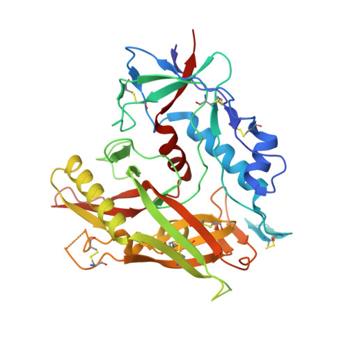
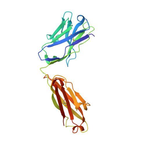
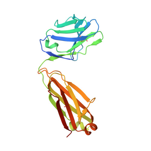
Identification of Near-Pan-neutralizing Antibodies against HIV-1 by Deconvolution of Plasma Humoral Responses.

Sajadi, M.M.Dashti, A.Rikhtegaran Tehrani, Z.Tolbert, W.D.Seaman, M.S.Ouyang, X.Gohain, N.Pazgier, M.Kim, D.Cavet, G.Yared, J.Redfield, R.R.Lewis, G.K.DeVico, A.L.

(2018) Cell 173: 1783-1795.e14

  • DOI: https://doi.org/10.1016/j.cell.2018.03.061
  • Primary Citation Related Structures: 
    5WB9, 6BCK

  • PubMed Abstract: 

    Anti-HIV-1 envelope broadly neutralizing monoclonal antibodies (bNAbs) isolated from memory B cells may not fully represent HIV-1-neutralizing profiles measured in plasma. Accordingly, we characterized near-pan-neutralizing antibodies extracted directly from the plasma of two "elite neutralizers." Circulating anti-gp120 polyclonal antibodies were deconvoluted using proteomics to guide lineage analysis of bone marrow plasma cells. In both subjects, a single lineage of anti-CD4-binding site (CD4bs) antibodies explained the plasma-neutralizing activity. Importantly, members of these lineages potently neutralized 89%-100% of a multi-tier 117 pseudovirus panel, closely matching the specificity and breadth of the circulating antibodies. X-ray crystallographic analysis of one monoclonal, N49P7, suggested a unique ability to bypass the CD4bs Phe43 cavity, while reaching deep into highly conserved residues of Layer 3 of the gp120 inner domain, likely explaining its extreme potency and breadth. Further direct analyses of plasma anti-HIV-1 bNAbs should provide new insights for developing antibody-based antiviral agents and vaccines.


  • Organizational Affiliation
    • Divisions of Vaccine Research and Clinical Care and Research, Institute of Human Virology, University of Maryland School of Medicine, Baltimore, MD 21201, USA; Department of Medicine, Baltimore VA Medical Center, Baltimore, MD 21201, USA; Department of Medicine, University of Maryland School of Medicine, Baltimore, MD 21201, USA. Electronic address: msajadi@ihv.umaryland.edu.

Macromolecule Content 

  • Total Structure Weight: 88.84 kDa 
  • Atom Count: 6,260 
  • Modeled Residue Count: 760 
  • Deposited Residue Count: 788 
  • Unique protein chains: 3

Macromolecules

Find similar proteins by:|  3D Structure
Entity ID: 1
MoleculeChains  Sequence LengthOrganismDetailsImage
clade A/E 93TH057 HIV-1 gp120 coreA [auth G]353Human immunodeficiency virus 1Mutation(s): 1 
Gene Names: HIV-1 Env
UniProt
Find proteins for A0A0M3KKW9 (Human immunodeficiency virus type 1)
Explore A0A0M3KKW9 
Go to UniProtKB:  A0A0M3KKW9
Entity Groups
Sequence Clusters30% Identity50% Identity70% Identity90% Identity95% Identity100% Identity
UniProt GroupA0A0M3KKW9
Glycosylation
Glycosylation Sites: 8
Sequence Annotations
Expand
Reference Sequence
Find similar proteins by:|  3D Structure
Entity ID: 2
MoleculeChains  Sequence LengthOrganismDetailsImage
N60P23 Fab heavy chainB [auth H]227Homo sapiensMutation(s): 0 
Find similar proteins by:|  3D Structure
Entity ID: 3
MoleculeChains  Sequence LengthOrganismDetailsImage
N60P23 Fab light chainC [auth L]208Homo sapiensMutation(s): 0 

Small Molecules

Ligands 3 Unique
IDChains Name / Formula / InChI Key2D Diagram3D Interactions
NAG

Query on NAG


Download:Ideal Coordinates CCD File
D [auth G]
E [auth G]
F [auth G]
G
H [auth G]
D [auth G],
E [auth G],
F [auth G],
G,
H [auth G],
I [auth G],
J [auth G],
K [auth G]
2-acetamido-2-deoxy-beta-D-glucopyranose
C8 H15 N O6
OVRNDRQMDRJTHS-FMDGEEDCSA-N
MPO

Query on MPO


Download:Ideal Coordinates CCD File
M [auth G]3[N-MORPHOLINO]PROPANE SULFONIC ACID
C7 H15 N O4 S
DVLFYONBTKHTER-UHFFFAOYSA-N
MPD

Query on MPD


Download:Ideal Coordinates CCD File
L [auth G](4S)-2-METHYL-2,4-PENTANEDIOL
C6 H14 O2
SVTBMSDMJJWYQN-YFKPBYRVSA-N

Experimental Data & Validation

Experimental Data

  • Method: X-RAY DIFFRACTION
  • Resolution: 2.40 Å
  • R-Value Free:  0.258 (Depositor), 0.259 (DCC) 
  • R-Value Work:  0.213 (Depositor), 0.217 (DCC) 
  • R-Value Observed: 0.216 (Depositor) 
Space Group: C 1 2 1
Unit Cell:
Length ( Å )Angle ( ˚ )
a = 127.562α = 90
b = 68.567β = 111.36
c = 119.356γ = 90
Software Package:
Software NamePurpose
REFMACrefinement
HKL-2000data reduction
HKL-2000data scaling
PHASERphasing

Structure Validation

View Full Validation Report


Ligand Structure Quality Assessment 


Entry History 

Deposition Data

Revision History  (Full details and data files)

  • Version 1.0: 2018-05-23
    Type: Initial release
  • Version 1.1: 2018-06-27
    Changes: Data collection, Database references
  • Version 1.2: 2020-07-29
    Type: Remediation
    Reason: Carbohydrate remediation
    Changes: Data collection, Derived calculations, Structure summary
  • Version 1.3: 2023-10-04
    Changes: Data collection, Database references, Refinement description, Structure summary
  • Version 1.4: 2024-11-13
    Changes: Structure summary