5WAI

Crystal Structure of a Suz12-Rbbp4-Jarid2-Aebp2 Heterotetrameric Complex


Experimental Data Snapshot

  • Method: X-RAY DIFFRACTION
  • Resolution: 2.90 Å
  • R-Value Free: 0.216 
  • R-Value Work: 0.171 
  • R-Value Observed: 0.173 

wwPDB Validation   3D Report Full Report


This is version 1.1 of the entry. See complete history


Literature

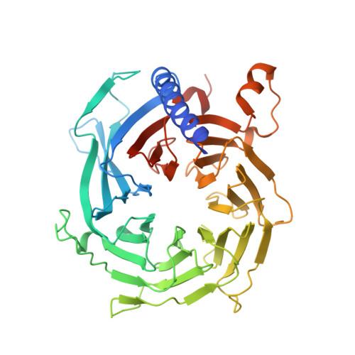
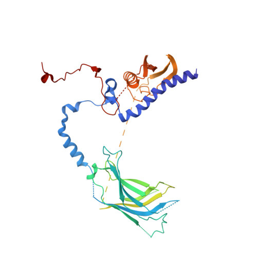
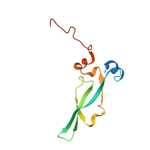
Unique Structural Platforms of Suz12 Dictate Distinct Classes of PRC2 for Chromatin Binding.

Chen, S.Jiao, L.Shubbar, M.Yang, X.Liu, X.

(2018) Mol Cell 69: 840-852.e5

  • DOI: https://doi.org/10.1016/j.molcel.2018.01.039
  • Primary Citation of Related Structures:  
    5WAI, 5WAK

  • PubMed Abstract: 

    Developmentally regulated accessory subunits dictate PRC2 function. Here, we report the crystal structures of a 120 kDa heterotetrameric complex consisting of Suz12, Rbbp4, Jarid2, and Aebp2 fragments that is minimally active in nucleosome binding and of an inactive binary complex of Suz12 and Rbbp4. Suz12 contains two unique structural platforms that define distinct classes of PRC2 holo complexes for chromatin binding. Aebp2 and Phf19 compete for binding of a non-canonical C2 domain of Suz12; Jarid2 and EPOP occupy an overlapped Suz12 surface required for chromatin association of PRC2. Suz12 and Aebp2 progressively block histone H3K4 binding to Rbbp4, suggesting that Rbbp4 may not be directly involved in PRC2 inhibition by the active H3K4me3 histone mark. Nucleosome binding enabled by Jarid2 and Aebp2 is in part accounted for by the structures, which also reveal that disruption of the Jarid2-Suz12 interaction may underlie the disease mechanism of an oncogenic chromosomal translocation of Suz12.


  • Organizational Affiliation

    Cecil H. and Ida Green Center for Reproductive Biology Sciences and Division of Basic Research, Department of Obstetrics and Gynecology, Department of Biophysics, UT Southwestern Medical Center, Dallas, TX 75390, USA.


Macromolecules
Find similar proteins by:  (by identity cutoff)  |  3D Structure
Entity ID: 1
MoleculeChains Sequence LengthOrganismDetailsImage
Histone-binding protein RBBP4
A, E
439Homo sapiensMutation(s): 0 
Gene Names: RBBP4RBAP48
UniProt & NIH Common Fund Data Resources
Find proteins for Q09028 (Homo sapiens)
Explore Q09028 
Go to UniProtKB:  Q09028
PHAROS:  Q09028
GTEx:  ENSG00000162521 
Entity Groups  
Sequence Clusters30% Identity50% Identity70% Identity90% Identity95% Identity100% Identity
UniProt GroupQ09028
Sequence Annotations
Expand
  • Reference Sequence
Find similar proteins by:  (by identity cutoff)  |  3D Structure
Entity ID: 2
MoleculeChains Sequence LengthOrganismDetailsImage
Polycomb protein SUZ12
B, F
478Homo sapiensMutation(s): 0 
Gene Names: SUZ12CHET9JJAZ1KIAA0160
UniProt & NIH Common Fund Data Resources
Find proteins for Q15022 (Homo sapiens)
Explore Q15022 
Go to UniProtKB:  Q15022
PHAROS:  Q15022
GTEx:  ENSG00000178691 
Entity Groups  
Sequence Clusters30% Identity50% Identity70% Identity90% Identity95% Identity100% Identity
UniProt GroupQ15022
Sequence Annotations
Expand
  • Reference Sequence
Find similar proteins by:  (by identity cutoff)  |  3D Structure
Entity ID: 3
MoleculeChains Sequence LengthOrganismDetailsImage
Zinc finger protein AEBP2
C, G
100Homo sapiensMutation(s): 0 
Gene Names: AEBP2
UniProt & NIH Common Fund Data Resources
Find proteins for Q6ZN18 (Homo sapiens)
Explore Q6ZN18 
Go to UniProtKB:  Q6ZN18
PHAROS:  Q6ZN18
GTEx:  ENSG00000139154 
Entity Groups  
Sequence Clusters30% Identity50% Identity70% Identity90% Identity95% Identity100% Identity
UniProt GroupQ6ZN18
Sequence Annotations
Expand
  • Reference Sequence

Find similar proteins by:  Sequence   |   3D Structure  

Entity ID: 4
MoleculeChains Sequence LengthOrganismDetailsImage
Jumonji, AT-rich interactive domain 2
D, H
19Neogale visonMutation(s): 0 
UniProt & NIH Common Fund Data Resources
Find proteins for Q92833 (Homo sapiens)
Explore Q92833 
Go to UniProtKB:  Q92833
PHAROS:  Q92833
GTEx:  ENSG00000008083 
Entity Groups  
Sequence Clusters30% Identity50% Identity70% Identity90% Identity95% Identity100% Identity
UniProt GroupQ92833
Sequence Annotations
Expand
  • Reference Sequence
Experimental Data & Validation

Experimental Data

  • Method: X-RAY DIFFRACTION
  • Resolution: 2.90 Å
  • R-Value Free: 0.216 
  • R-Value Work: 0.171 
  • R-Value Observed: 0.173 
  • Space Group: P 21 21 21
Unit Cell:
Length ( Å )Angle ( ˚ )
a = 95.3α = 90
b = 111.428β = 90
c = 253.254γ = 90
Software Package:
Software NamePurpose
BUSTERrefinement
HKL-2000data reduction
HKL-2000data scaling
PHASERphasing

Structure Validation

View Full Validation Report


Entry History 

Deposition Data

Revision History  (Full details and data files)

  • Version 1.0: 2018-03-14
    Type: Initial release
  • Version 1.1: 2023-10-04
    Changes: Data collection, Database references, Refinement description