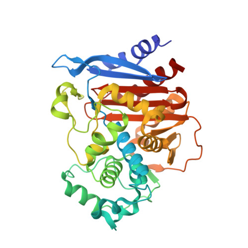
5WAC

ADC-7 in complex with boronic acid transition state inhibitor CR157


Experimental Data Snapshot

  • Method: X-RAY DIFFRACTION
  • Resolution: 2.06 Å
  • R-Value Free: 0.264 
  • R-Value Work: 0.203 
  • R-Value Observed: 0.206 

Starting Model: experimental
View more details

wwPDB Validation   3D Report Full Report


Ligand Structure Quality Assessment 


This is version 1.5 of the entry. See complete history


Literature

Structure-Based Analysis of Boronic Acids as Inhibitors of Acinetobacter-Derived Cephalosporinase-7, a Unique Class C beta-Lactamase.

Bouza, A.A.Swanson, H.C.Smolen, K.A.VanDine, A.L.Taracila, M.A.Romagnoli, C.Caselli, E.Prati, F.Bonomo, R.A.Powers, R.A.Wallar, B.J.

(2018) ACS Infect Dis 4: 325-336

  • DOI: https://doi.org/10.1021/acsinfecdis.7b00152
  • Primary Citation of Related Structures:  
    5WAC, 5WAD, 5WAE, 5WAF, 5WAG

  • PubMed Abstract: 

    Acinetobacter baumannii is a multidrug resistant pathogen that infects more than 12 000 patients each year in the US. Much of the resistance to β-lactam antibiotics in Acinetobacter spp. is mediated by class C β-lactamases known as Acinetobacter-derived cephalosporinases (ADCs). ADCs are unaffected by clinically used β-lactam-based β-lactamase inhibitors. In this study, five boronic acid transition state analog inhibitors (BATSIs) were evaluated for inhibition of the class C cephalosporinase ADC-7. Our goal was to explore the properties of BATSIs designed to probe the R1 binding site. K i values ranged from low micromolar to subnanomolar, and circular dichroism (CD) demonstrated that each inhibitor stabilizes the β-lactamase-inhibitor complexes. Additionally, X-ray crystal structures of ADC-7 in complex with five inhibitors were determined (resolutions from 1.80 to 2.09 Å). In the ADC-7/CR192 complex, the BATSI with the lowest K i (0.45 nM) and greatest Δ T m (+9 °C), a trifluoromethyl substituent, interacts with Arg340. Arg340 is unique to ADCs and may play an important role in the inhibition of ADC-7. The ADC-7/BATSI complexes determined in this study shed light into the unique recognition sites in ADC enzymes and also offer insight into further structure-based optimization of these inhibitors.


  • Organizational Affiliation

    Department of Chemistry , Grand Valley State University , 1 Campus Drive , Allendale , Michigan 49401 , United States.


Macromolecules
Find similar proteins by:  (by identity cutoff)  |  3D Structure
Entity ID: 1
MoleculeChains Sequence LengthOrganismDetailsImage
Beta-lactamase
A, B, C, D
361Acinetobacter baumanniiMutation(s): 0 
EC: 3.5.2.6
UniProt
Find proteins for Q6DRA1 (Acinetobacter baumannii)
Explore Q6DRA1 
Go to UniProtKB:  Q6DRA1
Entity Groups  
Sequence Clusters30% Identity50% Identity70% Identity90% Identity95% Identity100% Identity
UniProt GroupQ6DRA1
Sequence Annotations
Expand
  • Reference Sequence
Experimental Data & Validation

Experimental Data

  • Method: X-RAY DIFFRACTION
  • Resolution: 2.06 Å
  • R-Value Free: 0.264 
  • R-Value Work: 0.203 
  • R-Value Observed: 0.206 
  • Space Group: P 1 21 1
Unit Cell:
Length ( Å )Angle ( ˚ )
a = 88.352α = 90
b = 80.674β = 113.42
c = 104.98γ = 90
Software Package:
Software NamePurpose
REFMACrefinement
XDSdata reduction
XSCALEdata scaling
PHASERphasing
Cootmodel building

Structure Validation

View Full Validation Report


Ligand Structure Quality Assessment 


Entry History & Funding Information

Deposition Data


Funding OrganizationLocationGrant Number
National Institutes of Health/National Institute Of Allergy and Infectious Diseases (NIH/NIAID)United StatesAI072219

Revision History  (Full details and data files)

  • Version 1.0: 2017-12-06
    Type: Initial release
  • Version 1.1: 2017-12-20
    Changes: Database references
  • Version 1.2: 2018-03-21
    Changes: Database references
  • Version 1.3: 2019-12-11
    Changes: Author supporting evidence
  • Version 1.4: 2023-10-04
    Changes: Data collection, Database references, Refinement description
  • Version 1.5: 2024-10-23
    Changes: Structure summary