5W97

Crystal Structure of CO-bound Cytochrome c Oxidase determined by Serial Femtosecond X-Ray Crystallography at Room Temperature


Experimental Data Snapshot

  • Method: X-RAY DIFFRACTION
  • Resolution: 2.30 Å
  • R-Value Free: 0.252 
  • R-Value Work: 0.208 
  • R-Value Observed: 0.209 

Starting Model: experimental
View more details

wwPDB Validation   3D Report Full Report


Ligand Structure Quality Assessment 

Created with Raphaël 2.3.0Worse 01 BetterLigand structure goodness of fit to experimental dataBest fitted CDLClick on this verticalbar to view detailsBest fitted TGLClick on this verticalbar to view detailsBest fitted HEAClick on this verticalbar to view detailsBest fitted PEKClick on this verticalbar to view detailsBest fitted PSCClick on this verticalbar to view detailsBest fitted PGVClick on this verticalbar to view detailsBest fitted DMUClick on this verticalbar to view detailsBest fitted CHDClick on this verticalbar to view details

This is version 1.4 of the entry. See complete history


Literature

Crystal structure of CO-bound cytochrome c oxidase determined by serial femtosecond X-ray crystallography at room temperature.

Ishigami, I.Zatsepin, N.A.Hikita, M.Conrad, C.E.Nelson, G.Coe, J.D.Basu, S.Grant, T.D.Seaberg, M.H.Sierra, R.G.Hunter, M.S.Fromme, P.Fromme, R.Yeh, S.R.Rousseau, D.L.

(2017) Proc Natl Acad Sci U S A 114: 8011-8016

  • DOI: https://doi.org/10.1073/pnas.1705628114
  • Primary Citation of Related Structures:  
    5W97, 5WAU

  • PubMed Abstract: 

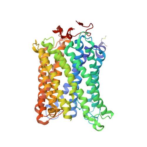
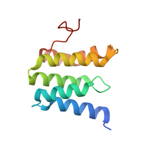
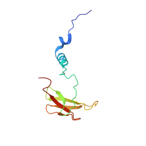
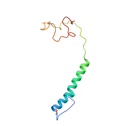
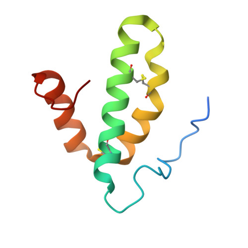
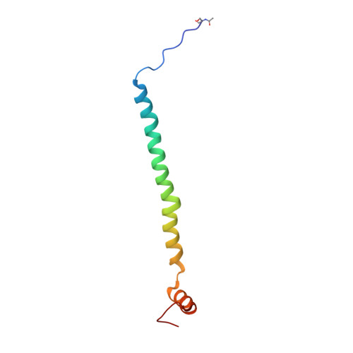
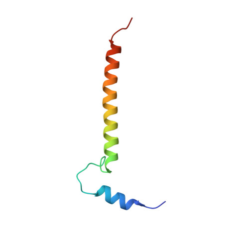
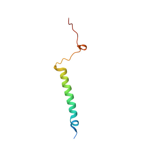
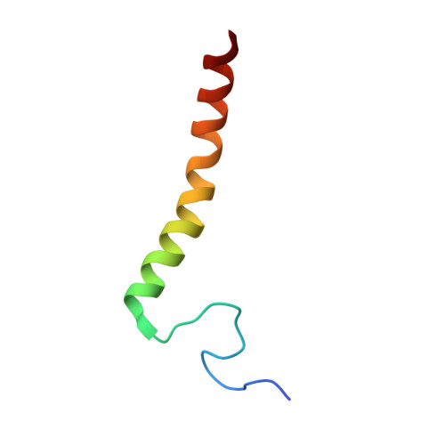
    Cytochrome c oxidase (C c O), the terminal enzyme in the electron transfer chain, translocates protons across the inner mitochondrial membrane by harnessing the free energy generated by the reduction of oxygen to water. Several redox-coupled proton translocation mechanisms have been proposed, but they lack confirmation, in part from the absence of reliable structural information due to radiation damage artifacts caused by the intense synchrotron radiation. Here we report the room temperature, neutral pH (6.8), damage-free structure of bovine C c O (bC c O) in the carbon monoxide (CO)-bound state at a resolution of 2.3 Å, obtained by serial femtosecond X-ray crystallography (SFX) with an X-ray free electron laser. As a comparison, an equivalent structure was obtained at a resolution of 1.95 Å, from data collected at a synchrotron light source. In the SFX structure, the CO is coordinated to the heme a 3 iron atom, with a bent Fe-C-O angle of ∼142°. In contrast, in the synchrotron structure, the Fe-CO bond is cleaved; CO relocates to a new site near Cu B , which, in turn, moves closer to the heme a 3 iron by ∼0.38 Å. Structural comparison reveals that ligand binding to the heme a 3 iron in the SFX structure is associated with an allosteric structural transition, involving partial unwinding of the helix-X between heme a and a 3 , thereby establishing a communication linkage between the two heme groups, setting the stage for proton translocation during the ensuing redox chemistry.


  • Organizational Affiliation

    Department of Physiology and Biophysics, Albert Einstein College of Medicine, Bronx, NY 10461.


Macromolecules
Find similar proteins by:  (by identity cutoff)  |  3D Structure
Entity ID: 1
MoleculeChains Sequence LengthOrganismDetailsImage
Cytochrome c oxidase subunit 1A,
N [auth a]
514Bos taurusMutation(s): 0 
EC: 1.9.3.1 (PDB Primary Data), 7.1.1.9 (UniProt)
Membrane Entity: Yes 
UniProt
Find proteins for P00396 (Bos taurus)
Explore P00396 
Go to UniProtKB:  P00396
Entity Groups  
Sequence Clusters30% Identity50% Identity70% Identity90% Identity95% Identity100% Identity
UniProt GroupP00396
Sequence Annotations
Expand
  • Reference Sequence
Find similar proteins by:  (by identity cutoff)  |  3D Structure
Entity ID: 2
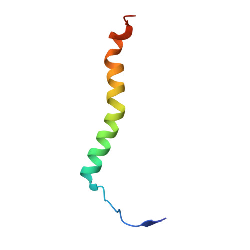
MoleculeChains Sequence LengthOrganismDetailsImage
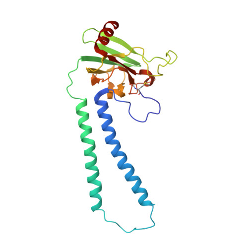
Cytochrome c oxidase subunit 2B,
O [auth b]
227Bos taurusMutation(s): 0 
EC: 7.1.1.9
Membrane Entity: Yes 
UniProt
Find proteins for P68530 (Bos taurus)
Explore P68530 
Go to UniProtKB:  P68530
Entity Groups  
Sequence Clusters30% Identity50% Identity70% Identity90% Identity95% Identity100% Identity
UniProt GroupP68530
Sequence Annotations
Expand
  • Reference Sequence
Find similar proteins by:  (by identity cutoff)  |  3D Structure
Entity ID: 3
MoleculeChains Sequence LengthOrganismDetailsImage
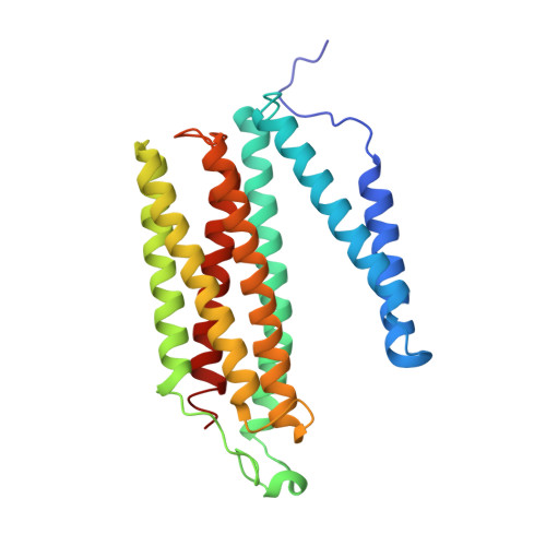
Cytochrome c oxidase subunit 3C,
P [auth c]
261Bos taurusMutation(s): 0 
EC: 7.1.1.9
Membrane Entity: Yes 
UniProt
Find proteins for P00415 (Bos taurus)
Explore P00415 
Go to UniProtKB:  P00415
Entity Groups  
Sequence Clusters30% Identity50% Identity70% Identity90% Identity95% Identity100% Identity
UniProt GroupP00415
Sequence Annotations
Expand
  • Reference Sequence
Find similar proteins by:  (by identity cutoff)  |  3D Structure
Entity ID: 4
MoleculeChains Sequence LengthOrganismDetailsImage
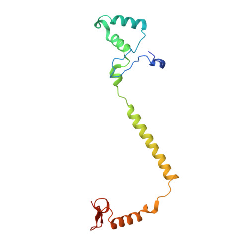
Cytochrome c oxidase subunit 4 isoform 1, mitochondrialD,
Q [auth d]
147Bos taurusMutation(s): 0 
Membrane Entity: Yes 
UniProt
Find proteins for P00423 (Bos taurus)
Explore P00423 
Go to UniProtKB:  P00423
Entity Groups  
Sequence Clusters30% Identity50% Identity70% Identity90% Identity95% Identity100% Identity
UniProt GroupP00423
Sequence Annotations
Expand
  • Reference Sequence
Find similar proteins by:  (by identity cutoff)  |  3D Structure
Entity ID: 5
MoleculeChains Sequence LengthOrganismDetailsImage
Cytochrome c oxidase subunit 5A, mitochondrialE,
R [auth e]
109Bos taurusMutation(s): 0 
Membrane Entity: Yes 
UniProt
Find proteins for P00426 (Bos taurus)
Explore P00426 
Go to UniProtKB:  P00426
Entity Groups  
Sequence Clusters30% Identity50% Identity70% Identity90% Identity95% Identity100% Identity
UniProt GroupP00426
Sequence Annotations
Expand
  • Reference Sequence
Find similar proteins by:  (by identity cutoff)  |  3D Structure
Entity ID: 6
MoleculeChains Sequence LengthOrganismDetailsImage
Cytochrome c oxidase subunit 5B, mitochondrialF,
S [auth f]
98Bos taurusMutation(s): 0 
Membrane Entity: Yes 
UniProt
Find proteins for P00428 (Bos taurus)
Explore P00428 
Go to UniProtKB:  P00428
Entity Groups  
Sequence Clusters30% Identity50% Identity70% Identity90% Identity95% Identity100% Identity
UniProt GroupP00428
Sequence Annotations
Expand
  • Reference Sequence
Find similar proteins by:  (by identity cutoff)  |  3D Structure
Entity ID: 7
MoleculeChains Sequence LengthOrganismDetailsImage
Cytochrome c oxidase subunit 6A2, mitochondrialG,
T [auth g]
85Bos taurusMutation(s): 0 
Membrane Entity: Yes 
UniProt
Find proteins for P07471 (Bos taurus)
Explore P07471 
Go to UniProtKB:  P07471
Entity Groups  
Sequence Clusters30% Identity50% Identity70% Identity90% Identity95% Identity100% Identity
UniProt GroupP07471
Sequence Annotations
Expand
  • Reference Sequence
Find similar proteins by:  (by identity cutoff)  |  3D Structure
Entity ID: 8
MoleculeChains Sequence LengthOrganismDetailsImage
Cytochrome c oxidase subunit 6B1H,
U [auth h]
85Bos taurusMutation(s): 0 
Membrane Entity: Yes 
UniProt
Find proteins for P00429 (Bos taurus)
Explore P00429 
Go to UniProtKB:  P00429
Entity Groups  
Sequence Clusters30% Identity50% Identity70% Identity90% Identity95% Identity100% Identity
UniProt GroupP00429
Sequence Annotations
Expand
  • Reference Sequence
Find similar proteins by:  (by identity cutoff)  |  3D Structure
Entity ID: 9
MoleculeChains Sequence LengthOrganismDetailsImage
Cytochrome c oxidase subunit 6CI,
V [auth i]
73Bos taurusMutation(s): 0 
Membrane Entity: Yes 
UniProt
Find proteins for P04038 (Bos taurus)
Explore P04038 
Go to UniProtKB:  P04038
Entity Groups  
Sequence Clusters30% Identity50% Identity70% Identity90% Identity95% Identity100% Identity
UniProt GroupP04038
Sequence Annotations
Expand
  • Reference Sequence
Find similar proteins by:  (by identity cutoff)  |  3D Structure
Entity ID: 10
MoleculeChains Sequence LengthOrganismDetailsImage
Cytochrome c oxidase subunit 7A1, mitochondrialJ,
W [auth j]
59Bos taurusMutation(s): 0 
Membrane Entity: Yes 
UniProt
Find proteins for P07470 (Bos taurus)
Explore P07470 
Go to UniProtKB:  P07470
Entity Groups  
Sequence Clusters30% Identity50% Identity70% Identity90% Identity95% Identity100% Identity
UniProt GroupP07470
Sequence Annotations
Expand
  • Reference Sequence
Find similar proteins by:  (by identity cutoff)  |  3D Structure
Entity ID: 11
MoleculeChains Sequence LengthOrganismDetailsImage
Cytochrome c oxidase subunit 7B, mitochondrialK,
X [auth k]
56Bos taurusMutation(s): 0 
Membrane Entity: Yes 
UniProt
Find proteins for P13183 (Bos taurus)
Explore P13183 
Go to UniProtKB:  P13183
Entity Groups  
Sequence Clusters30% Identity50% Identity70% Identity90% Identity95% Identity100% Identity
UniProt GroupP13183
Sequence Annotations
Expand
  • Reference Sequence
Find similar proteins by:  (by identity cutoff)  |  3D Structure
Entity ID: 12
MoleculeChains Sequence LengthOrganismDetailsImage
Cytochrome c oxidase subunit 7C, mitochondrialL,
Y [auth l]
47Bos taurusMutation(s): 0 
Membrane Entity: Yes 
UniProt
Find proteins for P00430 (Bos taurus)
Explore P00430 
Go to UniProtKB:  P00430
Entity Groups  
Sequence Clusters30% Identity50% Identity70% Identity90% Identity95% Identity100% Identity
UniProt GroupP00430
Sequence Annotations
Expand
  • Reference Sequence
Find similar proteins by:  (by identity cutoff)  |  3D Structure
Entity ID: 13
MoleculeChains Sequence LengthOrganismDetailsImage
Cytochrome c oxidase subunit 8B, mitochondrialM,
Z [auth m]
46Bos taurusMutation(s): 0 
Membrane Entity: Yes 
UniProt
Find proteins for P10175 (Bos taurus)
Explore P10175 
Go to UniProtKB:  P10175
Entity Groups  
Sequence Clusters30% Identity50% Identity70% Identity90% Identity95% Identity100% Identity
UniProt GroupP10175
Sequence Annotations
Expand
  • Reference Sequence
Small Molecules
Ligands 14 Unique
IDChains Name / Formula / InChI Key2D Diagram3D Interactions
CDL
Query on CDL

Download Ideal Coordinates CCD File 
QA [auth C],
RB [auth c],
XA [auth G],
XB [auth g]
CARDIOLIPIN
C81 H156 O17 P2
XVTUQDWPJJBEHJ-KZCWQMDCSA-L
TGL
Query on TGL

Download Ideal Coordinates CCD File 
AC [auth l]
BB [auth L]
HA [auth A]
IB [auth a]
VA [auth D]
TRISTEAROYLGLYCEROL
C57 H110 O6
DCXXMTOCNZCJGO-UHFFFAOYSA-N
HEA
Query on HEA

Download Ideal Coordinates CCD File 
AA [auth A],
BA [auth A],
DB [auth a],
EB [auth a]
HEME-A
C49 H56 Fe N4 O6
ZGGYGTCPXNDTRV-PRYGPKJJSA-L
PEK
Query on PEK

Download Ideal Coordinates CCD File 
NA [auth C]
PB [auth c]
TA [auth C]
UA [auth C]
WB [auth g]
(1S)-2-{[(2-AMINOETHOXY)(HYDROXY)PHOSPHORYL]OXY}-1-[(STEAROYLOXY)METHYL]ETHYL (5E,8E,11E,14E)-ICOSA-5,8,11,14-TETRAENOATE
C43 H78 N O8 P
ANRKEHNWXKCXDB-BHFWLYLHSA-N
PSC
Query on PSC

Download Ideal Coordinates CCD File 
KA [auth B],
UB [auth e]
(7R,17E,20E)-4-HYDROXY-N,N,N-TRIMETHYL-9-OXO-7-[(PALMITOYLOXY)METHYL]-3,5,8-TRIOXA-4-PHOSPHAHEXACOSA-17,20-DIEN-1-AMINIUM 4-OXIDE
C42 H81 N O8 P
JLPULHDHAOZNQI-AUSZDXHESA-O
PGV
Query on PGV

Download Ideal Coordinates CCD File 
FA [auth A]
GA [auth A]
JB [auth a]
NB [auth c]
OA [auth C]
(1R)-2-{[{[(2S)-2,3-DIHYDROXYPROPYL]OXY}(HYDROXY)PHOSPHORYL]OXY}-1-[(PALMITOYLOXY)METHYL]ETHYL (11E)-OCTADEC-11-ENOATE
C40 H77 O10 P
ADYWCMPUNIVOEA-GPJPVTGXSA-N
DMU
Query on DMU

Download Ideal Coordinates CCD File 
BC [auth m],
CB [auth M],
SA [auth C],
TB [auth c]
DECYL-BETA-D-MALTOPYRANOSIDE
C22 H42 O11
WOQQAWHSKSSAGF-WXFJLFHKSA-N
CHD
Query on CHD

Download Ideal Coordinates CCD File 
AB [auth J]
LA [auth B]
LB [auth b]
MA [auth C]
OB [auth c]
CHOLIC ACID
C24 H40 O5
BHQCQFFYRZLCQQ-OELDTZBJSA-N
CUA
Query on CUA

Download Ideal Coordinates CCD File 
JA [auth B],
MB [auth b]
DINUCLEAR COPPER ION
Cu2
ALKZAGKDWUSJED-UHFFFAOYSA-N
ZN
Query on ZN

Download Ideal Coordinates CCD File 
VB [auth f],
WA [auth F]
ZINC ION
Zn
PTFCDOFLOPIGGS-UHFFFAOYSA-N
CU
Query on CU

Download Ideal Coordinates CCD File 
CA [auth A],
FB [auth a]
COPPER (II) ION
Cu
JPVYNHNXODAKFH-UHFFFAOYSA-N
CMO
Query on CMO

Download Ideal Coordinates CCD File 
IA [auth A],
KB [auth a]
CARBON MONOXIDE
C O
UGFAIRIUMAVXCW-UHFFFAOYSA-N
MG
Query on MG

Download Ideal Coordinates CCD File 
DA [auth A],
GB [auth a]
MAGNESIUM ION
Mg
JLVVSXFLKOJNIY-UHFFFAOYSA-N
NA
Query on NA

Download Ideal Coordinates CCD File 
EA [auth A],
HB [auth a]
SODIUM ION
Na
FKNQFGJONOIPTF-UHFFFAOYSA-N
Modified Residues  3 Unique
IDChains TypeFormula2D DiagramParent
FME
Query on FME
A,
N [auth a]
L-PEPTIDE LINKINGC6 H11 N O3 SMET
TPO
Query on TPO
G,
T [auth g]
L-PEPTIDE LINKINGC4 H10 N O6 PTHR
SAC
Query on SAC
I,
V [auth i]
L-PEPTIDE LINKINGC5 H9 N O4SER
Experimental Data & Validation

Experimental Data

  • Method: X-RAY DIFFRACTION
  • Resolution: 2.30 Å
  • R-Value Free: 0.252 
  • R-Value Work: 0.208 
  • R-Value Observed: 0.209 
  • Space Group: P 21 21 21
Unit Cell:
Length ( Å )Angle ( ˚ )
a = 178.3α = 90
b = 189β = 90
c = 209γ = 90
Software Package:
Software NamePurpose
REFMACrefinement
CrystFELdata scaling
MOSFLMdata reduction
PHASERphasing

Structure Validation

View Full Validation Report


Ligand Structure Quality Assessment 

Created with Raphaël 2.3.0Worse 01 BetterLigand structure goodness of fit to experimental dataBest fitted CDLClick on this verticalbar to view detailsBest fitted TGLClick on this verticalbar to view detailsBest fitted HEAClick on this verticalbar to view detailsBest fitted PEKClick on this verticalbar to view detailsBest fitted PSCClick on this verticalbar to view detailsBest fitted PGVClick on this verticalbar to view detailsBest fitted DMUClick on this verticalbar to view detailsBest fitted CHDClick on this verticalbar to view details

Entry History & Funding Information

Deposition Data


Funding OrganizationLocationGrant Number
National Institutes of Health/National Institute of General Medical Sciences (NIH/NIGMS)United StatesGM098799
National Institutes of Health/National Institute of General Medical Sciences (NIH/NIGMS)United StatesGM115773
National Institutes of Health/National Institute of General Medical Sciences (NIH/NIGMS)United StatesGM095583
National Science Foundation (NSF, United States)United StatesSTC-1231306
National Science Foundation (NSF, United States)United StatesCHE-1404929
National Science Foundation (NSF, United States)United StatesABI-1565180
National Institutes of Health/National Institute of General Medical Sciences (NIH/NIGMS)United StatesP41GM103393
Department of Energy (DOE, United States)United StatesDE-AC02-76SF00515

Revision History  (Full details and data files)

  • Version 1.0: 2017-08-09
    Type: Initial release
  • Version 1.1: 2017-09-20
    Changes: Author supporting evidence
  • Version 1.2: 2017-09-27
    Changes: Data collection
  • Version 1.3: 2019-11-27
    Changes: Author supporting evidence
  • Version 1.4: 2023-10-04
    Changes: Data collection, Database references, Derived calculations, Refinement description, Structure summary