5W8F

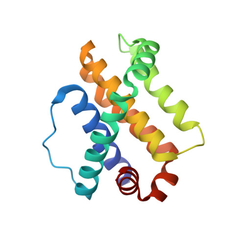
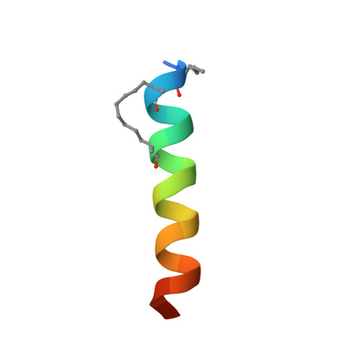
Crystal structure of human Mcl-1 in complex with modified Bim BH3 peptide SAH-MS1-14


Experimental Data Snapshot

  • Method: X-RAY DIFFRACTION
  • Resolution: 1.85 Å
  • R-Value Free: 0.289 
  • R-Value Work: 0.242 
  • R-Value Observed: 0.244 

Starting Model: experimental
View more details

wwPDB Validation   3D Report Full Report


This is version 2.1 of the entry. See complete history


Literature

Iterative optimization yields Mcl-1-targeting stapled peptides with selective cytotoxicity to Mcl-1-dependent cancer cells.

Rezaei Araghi, R.Bird, G.H.Ryan, J.A.Jenson, J.M.Godes, M.Pritz, J.R.Grant, R.A.Letai, A.Walensky, L.D.Keating, A.E.

(2018) Proc Natl Acad Sci U S A 115: E886-E895

  • DOI: https://doi.org/10.1073/pnas.1712952115
  • Primary Citation of Related Structures:  
    5W89, 5W8F

  • PubMed Abstract: 

    Bcl-2 family proteins regulate apoptosis, and aberrant interactions of overexpressed antiapoptotic family members such as Mcl-1 promote cell transformation, cancer survival, and resistance to chemotherapy. Discovering potent and selective Mcl-1 inhibitors that can relieve apoptotic blockades is thus a high priority for cancer research. An attractive strategy for disabling Mcl-1 involves using designer peptides to competitively engage its binding groove, mimicking the structural mechanism of action of native sensitizer BH3-only proteins. We transformed Mcl-1-binding peptides into α-helical, cell-penetrating constructs that are selectively cytotoxic to Mcl-1-dependent cancer cells. Critical to the design of effective inhibitors was our introduction of an all-hydrocarbon cross-link or "staple" that stabilizes α-helical structure, increases target binding affinity, and independently confers binding specificity for Mcl-1 over related Bcl-2 family paralogs. Two crystal structures of complexes at 1.4 Å and 1.9 Å resolution demonstrate how the hydrophobic staple induces an unanticipated structural rearrangement in Mcl-1 upon binding. Systematic sampling of staple location and iterative optimization of peptide sequence in accordance with established design principles provided peptides that target intracellular Mcl-1. This work provides proof of concept for the development of potent, selective, and cell-permeable stapled peptides for therapeutic targeting of Mcl-1 in cancer, applying a design and validation workflow applicable to a host of challenging biomedical targets.


  • Organizational Affiliation

    Department of Biology, Massachusetts Institute of Technology, Cambridge, MA 02139.


Macromolecules
Find similar proteins by:  (by identity cutoff)  |  3D Structure
Entity ID: 1
MoleculeChains Sequence LengthOrganismDetailsImage
Induced myeloid leukemia cell differentiation protein Mcl-1150Homo sapiensMutation(s): 0 
Gene Names: MCL1BCL2L3
UniProt & NIH Common Fund Data Resources
Find proteins for Q07820 (Homo sapiens)
Explore Q07820 
Go to UniProtKB:  Q07820
PHAROS:  Q07820
GTEx:  ENSG00000143384 
Entity Groups  
Sequence Clusters30% Identity50% Identity70% Identity90% Identity95% Identity100% Identity
UniProt GroupQ07820
Sequence Annotations
Expand
  • Reference Sequence

Find similar proteins by:  Sequence   |   3D Structure  

Entity ID: 2
MoleculeChains Sequence LengthOrganismDetailsImage
modified Bim BH3 peptide SAH-MS1-1422Homo sapiensMutation(s): 0 
Entity Groups  
Sequence Clusters30% Identity50% Identity70% Identity90% Identity95% Identity100% Identity
Sequence Annotations
Expand
  • Reference Sequence
Small Molecules
Modified Residues  2 Unique
IDChains TypeFormula2D DiagramParent
MK8
Query on MK8
B
L-PEPTIDE LINKINGC7 H15 N O2LEU
NLE
Query on NLE
B
L-PEPTIDE LINKINGC6 H13 N O2LEU
Experimental Data & Validation

Experimental Data

  • Method: X-RAY DIFFRACTION
  • Resolution: 1.85 Å
  • R-Value Free: 0.289 
  • R-Value Work: 0.242 
  • R-Value Observed: 0.244 
  • Space Group: P 41 21 2
Unit Cell:
Length ( Å )Angle ( ˚ )
a = 46.85α = 90
b = 46.85β = 90
c = 167.44γ = 90
Software Package:
Software NamePurpose
PHENIXrefinement
XSCALEdata scaling
PDB_EXTRACTdata extraction
XDSdata reduction
PHASERphasing

Structure Validation

View Full Validation Report


Entry History 

Deposition Data

Revision History  (Full details and data files)

  • Version 1.0: 2018-01-17
    Type: Initial release
  • Version 1.1: 2018-01-31
    Changes: Database references
  • Version 1.2: 2018-02-14
    Changes: Database references
  • Version 1.3: 2023-10-04
    Changes: Data collection, Database references, Derived calculations, Refinement description
  • Version 2.0: 2023-11-15
    Changes: Atomic model, Data collection
  • Version 2.1: 2024-11-20
    Changes: Structure summary