RCSB PDB - 5W7Y: Crystal Structure of FHA domain of human APLF in complex with XRCC1 monophosphorylated mutated peptide

 5W7Y

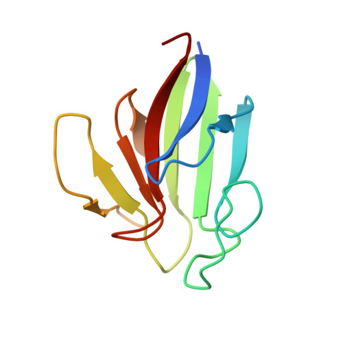
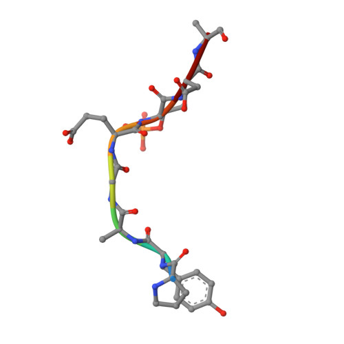
Crystal Structure of FHA domain of human APLF in complex with XRCC1 monophosphorylated mutated peptide


Experimental Data Snapshot

  • Method: X-RAY DIFFRACTION
  • Resolution: 2.10 Å
  • R-Value Free: 0.237 
  • R-Value Work: 0.203 
  • R-Value Observed: 0.204 

Starting Model: experimental
View more details

wwPDB Validation   3D Report Full Report


This is version 1.2 of the entry. See complete history


Literature

Characterization of the APLF FHA-XRCC1 phosphopeptide interaction and its structural and functional implications.

Kim, K.Pedersen, L.C.Kirby, T.W.DeRose, E.F.London, R.E.

(2017) Nucleic Acids Res 45: 12374-12387

  • DOI: https://doi.org/10.1093/nar/gkx941
  • Primary Citation of Related Structures:  
    5W7W, 5W7X, 5W7Y

  • PubMed Abstract: 

    Aprataxin and PNKP-like factor (APLF) is a DNA repair factor containing a forkhead-associated (FHA) domain that supports binding to the phosphorylated FHA domain binding motifs (FBMs) in XRCC1 and XRCC4. We have characterized the interaction of the APLF FHA domain with phosphorylated XRCC1 peptides using crystallographic, NMR, and fluorescence polarization studies. The FHA-FBM interactions exhibit significant pH dependence in the physiological range as a consequence of the atypically high pK values of the phosphoserine and phosphothreonine residues and the preference for a dianionic charge state of FHA-bound pThr. These high pK values are characteristic of the polyanionic peptides typically produced by CK2 phosphorylation. Binding affinity is greatly enhanced by residues flanking the crystallographically-defined recognition motif, apparently as a consequence of non-specific electrostatic interactions, supporting the role of XRCC1 in nuclear cotransport of APLF. The FHA domain-dependent interaction of XRCC1 with APLF joins repair scaffolds that support single-strand break repair and non-homologous end joining (NHEJ). It is suggested that for double-strand DNA breaks that have initially formed a complex with PARP1 and its binding partner XRCC1, this interaction acts as a backup attempt to intercept the more error-prone alternative NHEJ repair pathway by recruiting Ku and associated NHEJ factors.


  • Organizational Affiliation

    Genome Integrity and Structural Biology Laboratory, National Institute of Environmental Health Sciences, National Institutes of Health, Research Triangle Park, NC 27709, USA.


Macromolecules
Find similar proteins by:  (by identity cutoff)  |  3D Structure
Entity ID: 1
MoleculeChains Sequence LengthOrganismDetailsImage
Aprataxin and PNK-like factorA,
C [auth B]
108Homo sapiensMutation(s): 0 
Gene Names: APLFC2orf13PALFXIP1
EC: 4.2.99.18 (PDB Primary Data), 3.1 (UniProt)
UniProt & NIH Common Fund Data Resources
Find proteins for Q8IW19 (Homo sapiens)
Explore Q8IW19 
Go to UniProtKB:  Q8IW19
PHAROS:  Q8IW19
GTEx:  ENSG00000169621 
Entity Groups  
Sequence Clusters30% Identity50% Identity70% Identity90% Identity95% Identity100% Identity
UniProt GroupQ8IW19
Sequence Annotations
Expand
  • Reference Sequence

Find similar proteins by:  Sequence   |   3D Structure  

Entity ID: 2
MoleculeChains Sequence LengthOrganismDetailsImage
DNA repair protein XRCC1B [auth D],
D [auth C]
8Homo sapiensMutation(s): 1 
UniProt & NIH Common Fund Data Resources
Find proteins for P18887 (Homo sapiens)
Explore P18887 
Go to UniProtKB:  P18887
PHAROS:  P18887
GTEx:  ENSG00000073050 
Entity Groups  
UniProt GroupP18887
Sequence Annotations
Expand
  • Reference Sequence
Small Molecules
Modified Residues  1 Unique
IDChains TypeFormula2D DiagramParent
TPO
Query on TPO
B [auth D],
D [auth C]
L-PEPTIDE LINKINGC4 H10 N O6 PTHR
Experimental Data & Validation

Experimental Data

  • Method: X-RAY DIFFRACTION
  • Resolution: 2.10 Å
  • R-Value Free: 0.237 
  • R-Value Work: 0.203 
  • R-Value Observed: 0.204 
  • Space Group: C 1 2 1
Unit Cell:
Length ( Å )Angle ( ˚ )
a = 54.799α = 90
b = 31.072β = 97.84
c = 118.908γ = 90
Software Package:
Software NamePurpose
PHENIXrefinement
HKL-3000data reduction
HKL-3000data scaling
PHASERphasing

Structure Validation

View Full Validation Report


Entry History 

Deposition Data

Revision History  (Full details and data files)

  • Version 1.0: 2018-05-02
    Type: Initial release
  • Version 1.1: 2023-10-04
    Changes: Data collection, Database references, Refinement description
  • Version 1.2: 2024-10-16
    Changes: Structure summary