5W4L | pdb_00005w4l

Crystal structure of the non-neutralizing and ADCC-potent C11-like antibody N12-i3 in complex with HIV-1 clade A/E gp120, the CD4 mimetic M48U1, and the antibody N5-i5.


Experimental Data Snapshot

  • Method: X-RAY DIFFRACTION
  • Resolution: 2.92 Å
  • R-Value Free: 
    0.292 (Depositor), 0.291 (DCC) 
  • R-Value Work: 
    0.231 (Depositor), 0.232 (DCC) 
  • R-Value Observed: 
    0.234 (Depositor) 

Starting Model: experimental
View more details

wwPDB Validation 3D Report Full Report

Validation slider image for 5W4L

Ligand Structure Quality Assessment 


This is version 1.7 of the entry. See complete history

Literature

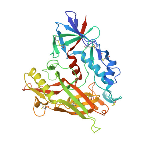
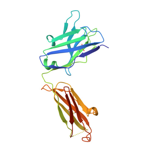
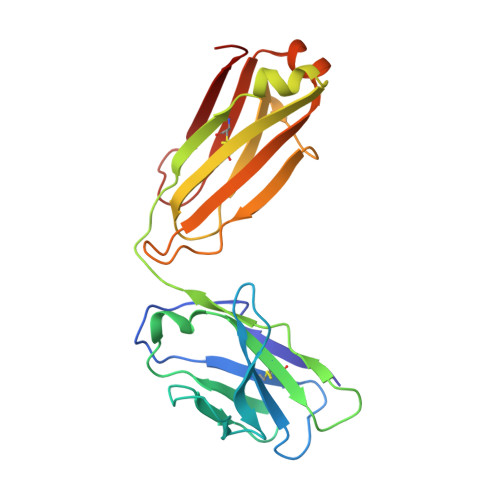
Targeting the Late Stage of HIV-1 Entry for Antibody-Dependent Cellular Cytotoxicity: Structural Basis for Env Epitopes in the C11 Region.

Tolbert, W.D.Gohain, N.Alsahafi, N.Van, V.Orlandi, C.Ding, S.Martin, L.Finzi, A.Lewis, G.K.Ray, K.Pazgier, M.

(2017) Structure 25: 1719-1731.e4

  • DOI: https://doi.org/10.1016/j.str.2017.09.009
  • Primary Citation Related Structures: 
    5W4L

  • PubMed Abstract: 

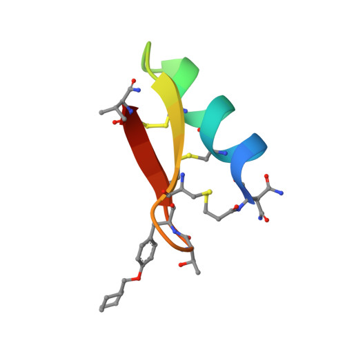
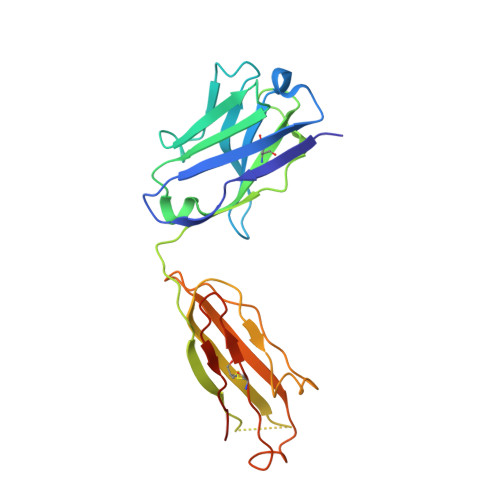
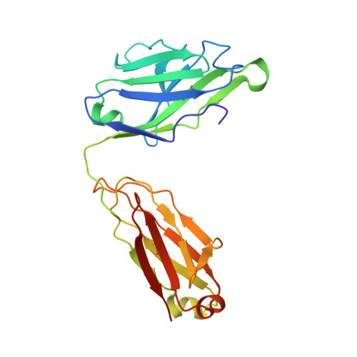
    Antibodies can have an impact on HIV-1 infection in multiple ways, including antibody-dependent cellular cytotoxicity (ADCC), a correlate of protection observed in the RV144 vaccine trial. One of the most potent ADCC-inducing epitopes on HIV-1 Env is recognized by the C11 antibody. Here, we present the crystal structure, at 2.9 Å resolution, of the C11-like antibody N12-i3, in a quaternary complex with the HIV-1 gp120, a CD4-mimicking peptide M48U1, and an A32-like antibody, N5-i5. Antibody N12-i3 recognizes an epitope centered on the N-terminal "eighth strand" of a critical β sandwich, which our analysis indicates to be emblematic of a late-entry state, after the gp120 detachment. In prior entry states, this sandwich comprises only seven strands, with the eighth strand instead pairing with a portion of the gp120 C terminus. The conformational gymnastics of HIV-1 gp120 thus includes altered β-strand pairing, possibly to reduce immunogenicity, although nevertheless still recognized by the human immune system.


  • Organizational Affiliation
    • Division of Vaccine Research, Institute of Human Virology, Baltimore, MD, USA; Department of Biochemistry and Molecular Biology, University of Maryland School of Medicine, 725 West Lombard Street, Baltimore, MD 21201, USA.

Macromolecule Content 

  • Total Structure Weight: 285.3 kDa 
  • Atom Count: 19,111 
  • Modeled Residue Count: 2,464 
  • Deposited Residue Count: 2,592 
  • Unique protein chains: 6

Macromolecules

Find similar proteins by:|  3D Structure
Entity ID: 1
MoleculeChains  Sequence LengthOrganismDetailsImage
clade A/E 93TH057 HIV-1 gp120 coreA [auth G],
G [auth A]
385Human immunodeficiency virus 1Mutation(s): 1 
Gene Names: HIV-1 Env
UniProt
Find proteins for A0A0M3KKW9 (Human immunodeficiency virus type 1)
Explore A0A0M3KKW9 
Go to UniProtKB:  A0A0M3KKW9
Entity Groups
Sequence Clusters30% Identity50% Identity70% Identity90% Identity95% Identity100% Identity
UniProt GroupA0A0M3KKW9
Glycosylation
Glycosylation Sites: 11
Sequence Annotations
Expand
Reference Sequence
Find similar proteins by:|  3D Structure
Entity ID: 2
MoleculeChains  Sequence LengthOrganismDetailsImage
CD4 mimetic peptide M48U1B [auth N],
H [auth M]
28synthetic constructMutation(s): 0 
Find similar proteins by:|  3D Structure
Entity ID: 3
MoleculeChains  Sequence LengthOrganismDetailsImage
Antibody N5-i5 Fab heavy chainC [auth H],
I [auth F]
226Homo sapiensMutation(s): 0 
Find similar proteins by:|  3D Structure
Entity ID: 4
MoleculeChains  Sequence LengthOrganismDetailsImage
Antibody N5-i5 light chainD [auth L],
J [auth I]
217Homo sapiensMutation(s): 0 
Find similar proteins by:|  3D Structure
Entity ID: 5
MoleculeChains  Sequence LengthOrganismDetailsImage
Antibody N12-i3 Fab heavy chainE [auth D],
K [auth B]
225Homo sapiensMutation(s): 0 
Find similar proteins by:|  3D Structure
Entity ID: 6
MoleculeChains  Sequence LengthOrganismDetailsImage
Antibody N12-i3 light chainF [auth E],
L [auth C]
215Homo sapiensMutation(s): 0 

Small Molecules

Ligands 2 Unique
IDChains Name / Formula / InChI Key2D Diagram3D Interactions
NAG

Query on NAG


Download:Ideal Coordinates CCD File
AA [auth A]
BA [auth A]
CA [auth A]
DA [auth A]
EA [auth A]
AA [auth A],
BA [auth A],
CA [auth A],
DA [auth A],
EA [auth A],
FA [auth A],
GA [auth A],
HA [auth A],
M [auth G],
N [auth G],
O [auth G],
P [auth G],
Q [auth G],
R [auth G],
S [auth G],
T [auth G],
U [auth G],
V [auth G],
W [auth G],
Y [auth A],
Z [auth A]
2-acetamido-2-deoxy-beta-D-glucopyranose
C8 H15 N O6
OVRNDRQMDRJTHS-FMDGEEDCSA-N
CL

Query on CL


Download:Ideal Coordinates CCD File
IA [auth A],
X [auth N]
CHLORIDE ION
Cl
VEXZGXHMUGYJMC-UHFFFAOYSA-M
Modified Residues  1 Unique
IDChains TypeFormula2D DiagramParent
DPR
Query on DPR
B [auth N],
H [auth M]
D-PEPTIDE LINKINGC5 H9 N O2

--

U2X
Query on U2X
B [auth N],
H [auth M]
L-PEPTIDE LINKINGC16 H23 N O3TYR

Biologically Interesting Molecules (External Reference) 

1 Unique

Experimental Data & Validation

Experimental Data

  • Method: X-RAY DIFFRACTION
  • Resolution: 2.92 Å
  • R-Value Free:  0.292 (Depositor), 0.291 (DCC) 
  • R-Value Work:  0.231 (Depositor), 0.232 (DCC) 
  • R-Value Observed: 0.234 (Depositor) 
Space Group: C 1 2 1
Unit Cell:
Length ( Å )Angle ( ˚ )
a = 311.018α = 90
b = 53.291β = 128.89
c = 223.577γ = 90
Software Package:
Software NamePurpose
REFMACrefinement
HKL-2000data reduction
HKL-2000data scaling
PHASERphasing

Structure Validation

View Full Validation Report


Ligand Structure Quality Assessment 


Entry History 

& Funding Information

Deposition Data


Funding OrganizationLocationGrant Number
National Institutes of Health/National Institute Of Allergy and Infectious Diseases (NIH/NIAID)United StatesAI116274

Revision History  (Full details and data files)

  • Version 1.0: 2017-11-15
    Type: Initial release
  • Version 1.1: 2017-12-06
    Changes: Author supporting evidence, Derived calculations
  • Version 1.2: 2018-04-18
    Changes: Data collection
  • Version 1.3: 2018-05-30
    Changes: Data collection, Structure summary
  • Version 1.4: 2019-12-11
    Changes: Author supporting evidence
  • Version 1.5: 2020-07-29
    Type: Remediation
    Reason: Carbohydrate remediation
    Changes: Data collection, Derived calculations, Structure summary
  • Version 1.6: 2023-10-04
    Changes: Data collection, Database references, Refinement description, Structure summary
  • Version 1.7: 2023-11-15
    Changes: Data collection