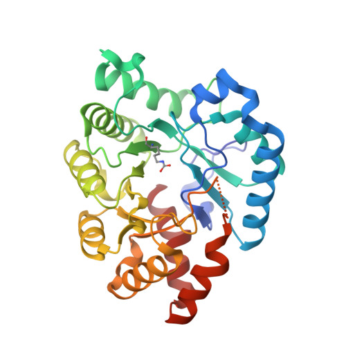
5W3Z

Crystal structure of SsoPox AsC6 mutant (L72I-Y99F-I122L-L228M-F229S-W263L)


Experimental Data Snapshot

  • Method: X-RAY DIFFRACTION
  • Resolution: 2.55 Å
  • R-Value Free: 0.252 
  • R-Value Work: 0.189 
  • R-Value Observed: 0.192 

Starting Model: experimental
View more details

wwPDB Validation   3D Report Full Report


This is version 2.2 of the entry. See complete history


Literature

Rational engineering of a native hyperthermostable lactonase into a broad spectrum phosphotriesterase.

Jacquet, P.Hiblot, J.Daude, D.Bergonzi, C.Gotthard, G.Armstrong, N.Chabriere, E.Elias, M.

(2017) Sci Rep 7: 16745-16745

  • DOI: https://doi.org/10.1038/s41598-017-16841-0
  • Primary Citation of Related Structures:  
    5VRI, 5VRK, 5VSA, 5W3U, 5W3W, 5W3Z

  • PubMed Abstract: 

    The redesign of enzyme active sites to alter their function or specificity is a difficult yet appealing challenge. Here we used a structure-based design approach to engineer the lactonase SsoPox from Sulfolobus solfataricus into a phosphotriesterase. The five best variants were characterized and their structure was solved. The most active variant, αsD6 (V27A-Y97W-L228M-W263M) demonstrates a large increase in catalytic efficiencies over the wild-type enzyme, with increases of 2,210-fold, 163-fold, 58-fold, 16-fold against methyl-parathion, malathion, ethyl-paraoxon, and methyl-paraoxon, respectively. Interestingly, the best mutants are also capable of degrading fensulfothion, which is reported to be an inhibitor for the wild-type enzyme, as well as others that are not substrates of the starting template or previously reported W263 mutants. The broad specificity of these engineered variants makes them promising candidates for the bioremediation of organophosphorus compounds. Analysis of their structures reveals that the increase in activity mainly occurs through the destabilization of the active site loop involved in substrate binding, and it has been observed that the level of disorder correlates with the width of the enzyme specificity spectrum. This finding supports the idea that active site conformational flexibility is essential to the acquisition of broader substrate specificity.


  • Organizational Affiliation

    CNRS UMR 7278, IRD198, INSERM U1095, APHM, Institut Hospitalier Universitaire Méditerranée-Infection, Aix-Marseille Université, 19-21 Bd Jean Moulin, 13005, Marseille, France.


Macromolecules
Find similar proteins by:  (by identity cutoff)  |  3D Structure
Entity ID: 1
MoleculeChains Sequence LengthOrganismDetailsImage
Aryldialkylphosphatase
A, B, C, D
314Saccharolobus solfataricusMutation(s): 6 
EC: 3.1.8.1
UniProt
Find proteins for Q97VT7 (Saccharolobus solfataricus (strain ATCC 35092 / DSM 1617 / JCM 11322 / P2))
Explore Q97VT7 
Go to UniProtKB:  Q97VT7
Entity Groups  
Sequence Clusters30% Identity50% Identity70% Identity90% Identity95% Identity100% Identity
UniProt GroupQ97VT7
Sequence Annotations
Expand
  • Reference Sequence
Small Molecules
Ligands 4 Unique
IDChains Name / Formula / InChI Key2D Diagram3D Interactions
GOL
Query on GOL

Download Ideal Coordinates CCD File 
H [auth A],
L [auth B],
Q [auth C],
U [auth D]
GLYCEROL
C3 H8 O3
PEDCQBHIVMGVHV-UHFFFAOYSA-N
EDO
Query on EDO

Download Ideal Coordinates CCD File 
G [auth A]
K [auth B]
O [auth C]
P [auth C]
T [auth D]
G [auth A],
K [auth B],
O [auth C],
P [auth C],
T [auth D],
V [auth D]
1,2-ETHANEDIOL
C2 H6 O2
LYCAIKOWRPUZTN-UHFFFAOYSA-N
CO
Query on CO

Download Ideal Coordinates CCD File 
F [auth A],
J [auth B],
N [auth C],
S [auth D]
COBALT (II) ION
Co
XLJKHNWPARRRJB-UHFFFAOYSA-N
FE2
Query on FE2

Download Ideal Coordinates CCD File 
E [auth A],
I [auth B],
M [auth C],
R [auth D]
FE (II) ION
Fe
CWYNVVGOOAEACU-UHFFFAOYSA-N
Modified Residues  1 Unique
IDChains TypeFormula2D DiagramParent
KCX
Query on KCX
A, B, C, D
L-PEPTIDE LINKINGC7 H14 N2 O4LYS
Experimental Data & Validation

Experimental Data

  • Method: X-RAY DIFFRACTION
  • Resolution: 2.55 Å
  • R-Value Free: 0.252 
  • R-Value Work: 0.189 
  • R-Value Observed: 0.192 
  • Space Group: P 21 21 21
Unit Cell:
Length ( Å )Angle ( ˚ )
a = 81.65α = 90
b = 105.8β = 90
c = 154.53γ = 90
Software Package:
Software NamePurpose
REFMACrefinement
XDSdata reduction
XSCALEdata scaling
REFMACphasing

Structure Validation

View Full Validation Report


Entry History 

Deposition Data

Revision History  (Full details and data files)

  • Version 1.0: 2018-01-10
    Type: Initial release
  • Version 1.1: 2019-11-20
    Changes: Derived calculations
  • Version 2.0: 2021-08-18
    Changes: Advisory, Atomic model, Data collection, Database references, Derived calculations, Non-polymer description, Structure summary
  • Version 2.1: 2023-10-04
    Changes: Data collection, Refinement description
  • Version 2.2: 2023-11-15
    Changes: Data collection